 ANSIR Processing Pipeline
 The Advanced NeuroScience Imaging Research lab (ANSIR) is devoted to the application of novel image analysis methods (e.g. diffeomorphic registration, machine learning, graph theory, ASL) to research studies, as well as to robust clinical translation of these techniques.
These methods have been applied to various studies on the brain, including: Diabetes, Dyslexia, Caffeine, Aging, Music, Alzheimer’s disease, Brain Tumors, Traumatic Brain Injury, and Neonatal Development. The ANSIR lab also maintains a fully automated functional and structural image processing pipeline supporting the image storage and analysis needs of a variety of scientists and imaging studies.
Methods
Arterial Spin Label (ASL)
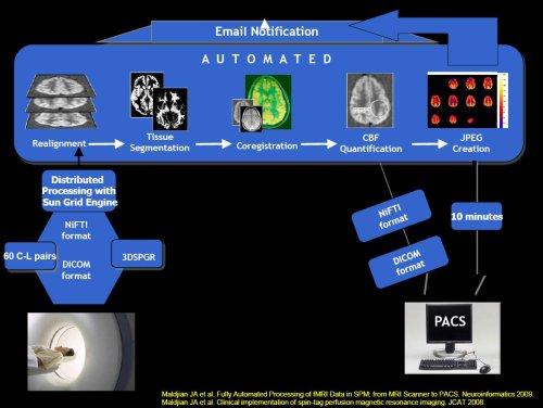
Arterial Spin Label MRI is a noninvasive method for measuring perfusion. Unlike traditional contrast, this method is completely non-invasive, is repeatable, and provides quantitative measures of blood flow. The ANSIR lab is developing techniques for ASL research and clinical applications.
Diffusion Tensor Imaging (DTI)
Diffusion Tensor Imaging uses MRI to measure the microscopic movement of water molecules. It provides measures of white matter axonal integrity and can be used in research studies of diseased and normal states, as well as for visualization of white matter tracts for clinical neurosurgical guidance.
Functional MRI (FMRI)
Functional MRI uses changes in local blood oxygen content to identify areas in the brain involved in performance of specific tasks. Resting state FMRI can be used to identify underlying brain networks, as well as changes related to disease or state-based modification of brain connectivity.
Structural MRI
Structural MRI analysis methods use high-resolution volumetric MRI data to identify differences in brain structure (e.g., grey matter, cortical thickness) between individuals or groups of subjects.
Magnetoencephalography (MEG)
Magnetoencephalography (MEG) is a non-invasive imaging technique that measures the magnetic fields produced by the electrical currents associated with neuronal function. It provides extremely high temporal resolution, and is complementary to structural and functional MRI methods. The ANSIR lab has developed a high-throughput fully automated MEG analysis pipeline for high density source-space analysis of resting state MEG data, incorporating a variety of head models, forward models, beamformers, and connectivity metrics.
Machine Learning
Machine learning has emerged as a powerful method of performing spatial pattern analysis and data classification. The multivariate nature of these approaches allows them to take into consideration correlations present in the data, overcoming limitations of standard analytical approaches. The prediction capabilities of machine learning methods are ideal for many clinical applications.
Network Analysis
Graph Theory based analysis of brain imaging data models the brain as a complex network represented as a collection of nodes and connecting edges. Nodes are typically defined as voxels (or regions of interest) in imaging space, and the edges are identified based on some connectivity measure between nodes (e.g. correlation coefficient). This framework allows the application of a variety of graph-theory based metrics to brain imaging data in order to identify and understand differences in brain connectivity between groups. While this has typically been applied to resting state fMRI data, graph theoretic methods can also be used with structural imaging data (e.g. DTI, as well as T1-weighted MRI).