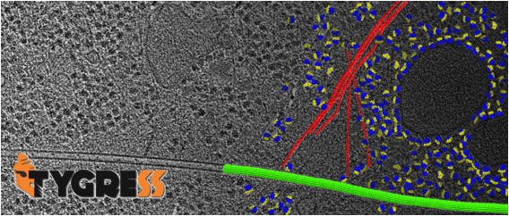
Tomography-Guided 3D Reconstruction of Subcellular Structures (TYGRESS) is a hybrid cellular-imaging method that combines the advantageous features of both cryo-electron tomography and single-particle cryo-EM to achieve higher resolution than before of complex subcellular structures within their native environment.
In this method, first an image is recorded with an electron dose that is typical for single-particle cryo-EM (e.g. 30 e-/Å2), followed by a traditional cryo-ET tilt series where for each image a much lower dose is applied (1-2 e-/Å2). The tomographic reconstruction and subtomogram reconstruction is used to guide the particle picking in the single-particle cryo-EM image and initial particle alignment.
With this guiding information, single-particle reconstruction and image-processing techniques can be applied to relatively thick cellular samples for the first time.
For support and questions, please contact Dr. Zhiguo Shang via Email
Downloads
- TYGRESS code
- Beta Version 1.0 Beta Development Version of TYGRESS: This beta version of TYGRESS has been released to and successfully tested by our local users. However, testing by external users has just begun. You might encounter bugs during the installation in your particular computer environment or while running the package. We greatly appreciate your reporting such bugs to Zhiguo Shang as we continue developing a first generally released version of TYGRESS.
- Version 1.1
- User manual
- Sample data set (coming soon)
References
- Z. Shang, X. Fu, K. Song, N. Grigorieff, D. Nicastro, Cellular imaging with improve resolution using TYGRESS: TomographY-Guided 3D REconstruction of Subcellular Structures. (in preparation)
- K. Song, Z. Shang, M. Carlone, D. Nicastro, TYGRESS reconstruction of the ciliary axoneme with up to 12 angstrom resolution. (in preparation)