Acquisition and Analysis of Human Neurophysiological Signals
Electroencephalography (EEG) – EEG is a noninvasive technique that can be used to record brain waves in both normal subjects and people with a particular disease. We not only record spontaneous activity, but can record signals while our research participants perform very specific behavioral tasks. Through a collaboration, we also record wireless mobile EEG signals to understand how brain signals change during complex movements, like walking, with a special interest in understanding why people with certain diagnoses have abnormal gait.
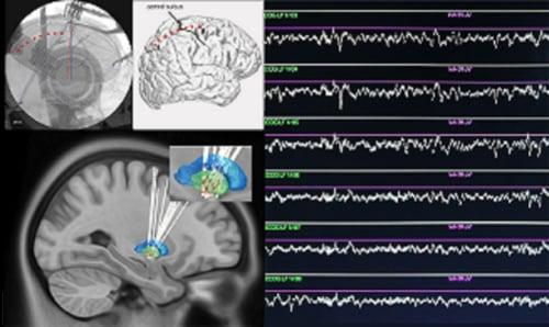
Electrocorticography (ECoG) – ECoG involves putting recording electrodes directly on the surface of the brain, rather than recording brain signals non-invasively with EEG. Because the scalp and skull are not in the way, the signals are much richer, with greater spectral content and greater spatial specificity. This allows us to explore very specific questions about the spatial localization of brain activity and relate activity across different parts of the brain. Analyses of these signals include spectral power, inter and intraregional functional connectivity (such as coherence and phase based connectivity, phase and amplitude cross frequency coupling), and other machine learning algorithms.
Local Field Potential (LFP) - LFP are similar to ECoG, in that they are recorded invasively, but instead of using surface electrodes, we record signals from deep within the brain using penetrating electrodes. In our lab, we most often use the deep brain stimulator leads that we are already implanting for research purposes. The same analyses used for ECoG can and are applied to LFPs.
Analysis of Kinematics of Movement
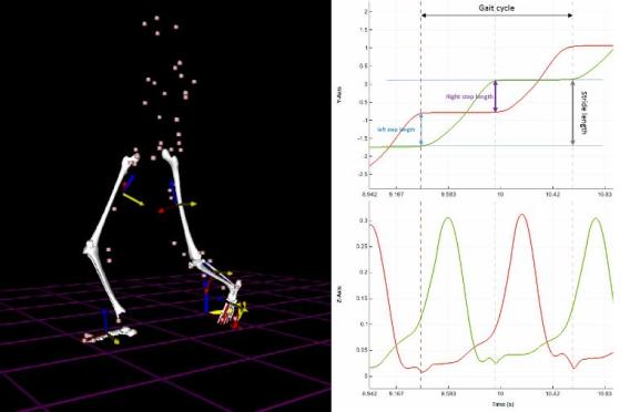
Movement is a very complex process, with subtle coordination and timing of different parts of the body. As part of our work, we are interested in understanding how the brain coordinates such complex movements, such as gait and walking. We use both a gait laboratory and multi-limb motion sensors to instantaneously record and stream movement details with synchronized neural signals. This data helps us to perform high temporal resolution comparisons between brain activity and movement. This research aims to understand “what goes wrong” in disease and identify brain regions, signals, or events that can be targeted for therapy.
Diffusion Tractography
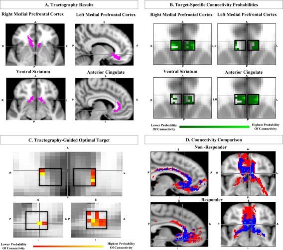
One of the core tenets of our lab is that the brain is a network – and that both disease and therapies affect networks. Therefore, we harness the power of advanced imaging to study these networks. Diffusion tractography is a non-invasive method of analyzing structural brain networks. Different parts of the brain are connected with white matter – which are essentially bundles of wires that interconnect the brain. Because water tends to move in the same direction as those “wires”, we can use a special MRI technique called diffusion imaging to measure that water movement. Those images can then be analyzed to identify structural connections between different parts of the brain. We have used these advanced imaging techniques to develop new ways of targeting brain stimulation, to understand the underlying cause of various symptoms within a disease, and to understand why some people respond to some therapies while others do not.
Resting and TaskBased Functional MRI
Besides structural connections in the brain, we can also use MRI to understand how different parts of the brain are functionally connected. Using functional MRI, we can measure brain activity in every pixel of the brain and then determine which parts of the brain are acting in similar manners. By looking at similarity in activity, we presume that 2 areas are “functionally connected.” We can also do functional MRI while subjects perform tasks in the MRI scanner, and have recently developed techniques (in collaboration with a team at Cleveland Clinic) to perform functional MRI in patients with deep brain stimulators. These functional MRI methods allow us to study differences in the functional brain architecture of people with disease compared to healthy controls as well as provides a unique opportunity to understand how treatment like deep brain stimulation affect brain networks and lead to improvements in symptoms.
Join Our Lab
Learn about opportunities to work with the Pouratian Lab.