We are actively developing high-performance attenuation correction strategies that utilize the PET signal alone.
Quantitative tracer images from combined PET-CT and PET-MR are increasingly being utilized for radiological decision-making and clinical trials. CT and MR based data corrections for PET photon attenuation, typically the largest impact on tracer quantification, can lead to increased radiation dose to the subject or require lengthy additional dedicated MR scans, respectively. Reconstruction methods that jointly estimate both the measured attenuation and tracer contrast from the subject PET signal represent a viable alternative, but, to date, have failed to match the quantification of CT approaches. Consequently, no single approach for PET attenuation correction that consistently produces high quantification and does not compromise patient safety or throughput currently exists. This is an important problem, since research and clinical findings must balance these practical issues with data quality.
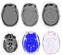
One area we are exploring are new acquisition and reconstruction strategies for attenuation correction that leverage the additional information from an external source. This work is being funded in part by an NIH NIBIB R03. For PET-CT these methods will benefit exams where the CT is used only for PET data corrections while for PET-MR the methods may improve tracer quantification where MR has limited performance and increase patient throughput by eliminating the need for non-diagnostic attenuation-only MR acquisitions. Particularly for brain imaging (image above) the proposed method is expected to greatly improve quantification compared to algorithms that use the signal from the patient alone.