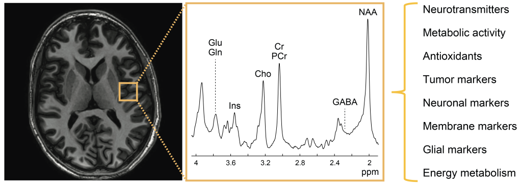
Magnetic resonance spectroscopy (MRS)
Magnetic resonance spectroscopy (MRS) is a powerful, noninvasive tool that provides a window into the biochemical processes in the brain without the need for ionizing radiation.
The Chemical Advanced Neuroimaging (CHAN) lab is committed to developing state-of-the-art proton MRS and MRS imaging (MRSI) methods to facilitate the techniques' translation to clinical practice. We also leverage these tools to answer key biological questions in clinical neuroscience and improve the diagnosis, disease monitoring, and treatment planning of a variety of neurological diseases and disorders, thus lengthening life and reducing illness and disability. Specific focus areas are detailed below.
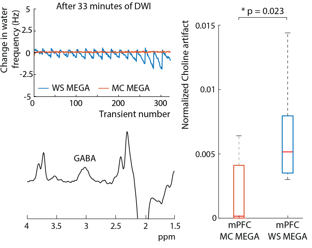
The newly developed MC MEGA method is robust to low-frequency offsets and choline artifacts, both of which are typically caused by experimental imperfections.
J-difference editing methods
Although in vivo proton MRS allows for the detection of a large range of neurochemicals essential to brain function, it suffers from high spectral overlap. Thus, specialized methods such as J-difference editing are typically used to detect low-concentration metabolites such as GABA, the brain’s main inhibitory neurotransmitter. A major focus of our lab is overcoming current technical barriers and improving the applicability of J-difference editing MRS methods to biomedical research and clinical practice. These technical advances include: (1) accelerating acquisitions (2) improving detection and (3) improving quantification. We also work with national and international collaborators to use our methods to study different diseases and disorders affecting the brain, such as cerebral palsy and gliomas.
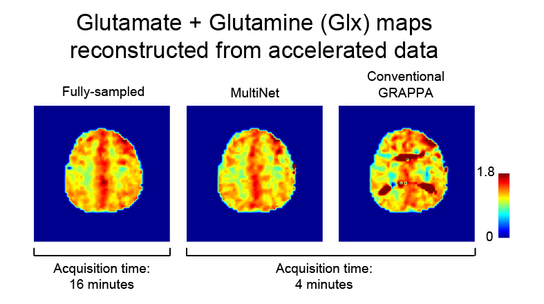
The GIx maps reconstructed with the novel MultiNet method is more similar to that of the fully sampled data than the GIx maps reconstructed with conventional GRAPPA.
Multi-voxel methods
While single-voxel 1H-MRS methods are conventionally used, these methods have limited spatial coverage. Thus, multi-voxel techniques are needed when multiple regions are of interest or if the affected region is unknown. These techniques have some limitations, however, including a long acquisition duration and sensitivity to motion. As such, a major focus of our lab is on overcoming some of these limitations by (1) accelerating acquisitions and (2) compensating for motion.
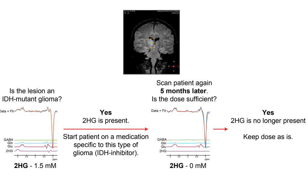
Using MRS to diagnose and monitor the treatment of IDH-mutant gliomas. Patient with lesion in the thalamus which cannot be diagnosed via biopsy without causing neurological injury.
Gliomas
Gliomas are brain tumors of glial origin and are the most common type of primary brain tumors. Mutations in the IDH1 and IDH2 genes are also present in over 70% of low-grade gliomas and are associated with a better prognosis compared to tumors without the IDH mutation. These mutations also result in the excess production of 2HG, which is detectable with MRS. As such, a major focus of our lab is the development and clinical translation of next-generation 2HG MRS methods as diagnostic, prognostic, and treatment planning tools for precision oncology. We also seek to better understand the metabolic changes that result from these genetic changes, which will help in the development of highly-effective glioma treatments.
Mood disorders
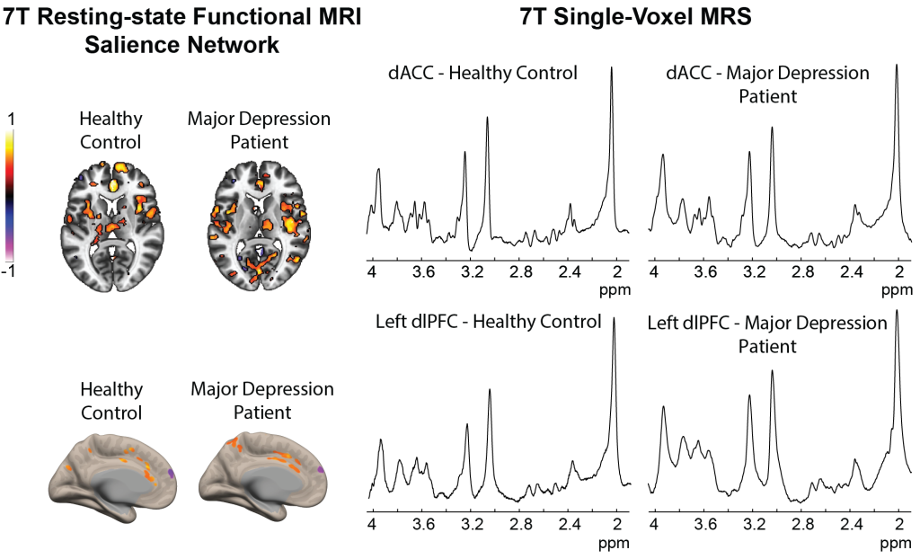
Bipolar disorder and major depressive disorder are two of the most common mood disorders with significant personal, societal, and economic burden. Our group is working on examining the relationship between changes in the neurotransmitters GABA and glutamate and changes in functional connectivity as measured by MRS and resting-state functional MRI, respectively, to better understand the biological mechanisms underlying functional changes in both disorders. By doing so, we hope to better understand the pathophysiology of both disorders which will help with developing better methods of diagnosing and treating these patients.