Metabolic MRI
Metabolic information of the human brain, body and skeletal muscle can be obtained by in vivo magnetic resonance spectroscopy (MRS), magnetic resonance spectroscopic imaging (MRSI), non-proton MRI and MRS as well as chemical exchange saturation transfer (CEST) imaging. While most of these methods use endogenous contrasts, some need simplistic tracers such as 13C or 2H labeled glucose.
1H MRS & MRSI
Proton magnetic resonance spectroscopy (1H MRS) and spectroscopic imaging (1H MRSI) are non-invasive methods that allow for the detection of about 20 metabolites and compounds in the brain and body without need of contrast agents. It provides rich information about energy metabolism, neurotransmission, myelination, cell membrane metabolism, osmoregulation, antioxidant profile and protein synthesis. 1H MRS can be applied for human as well as preclinical studies. The investigation of steady state mean tissue concentrations as well as their dynamic changes is possible.
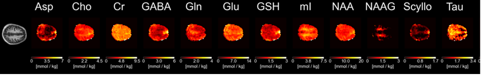
Adapted from: Wright AM, Murali-Manohar S, Henning A. Quantitative T1-relaxation corrected metabolite mapping of 12 metabolites in the human brain at 9.4 T. NeuroImage 263, 119574 (2022).
31P MRS & MRSI
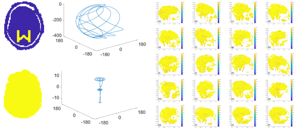
Phosphorous magnetic resonance spectroscopy (31P MRS) and spectroscopic imaging (31P MRSI) are non-invasive methods that allows for the detection of about 10 metabolites and compounds in the brain and body without need of contrast agents. It provides rich information about energy metabolism and cell membrane metabolisms and allows to determine intracellular and extracellular pH, magnesium concentrations and metabolic turnover rates. The latter is possible via saturation transfer experiments and via in bore exercise. 31P MRS can be applied for human as well as preclinical studies. The investigation of steady state mean tissue concentrations as well as their dynamic changes is possible.
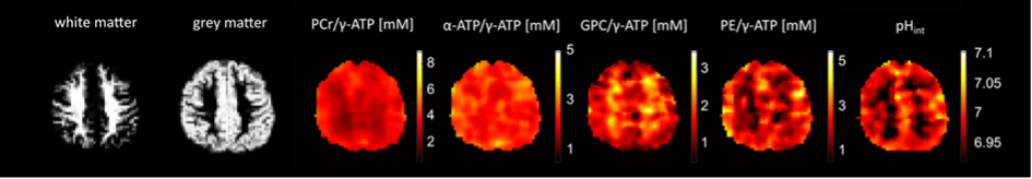
Ruhm L, Dorst J, Avdievitch N, Wright AM, Henning A. 3D 31P Magnetic Resonance Spectroscopic Imaging of the Human Brain at 9.4 Tesla: Optimization and Quantitative analysis of metabolic images. Magnetic Resonance in Medicine, 86 (5), pp. 2368 - 2383 (2021).
13C MRS & MRSI
Carbon magnetic resonance spectroscopy (13C MRS) is a non-invasive method that allows for the detection and detailed characterization of fatty acids and glycogen based on the 1% natural abundance of the MRI detectable isotope 13C. Furthermore, the enrichment of 13C labeling can be observed in about 10 additional metabolites and compounds in the brain and body after intake of 13C labeled glucose. Respective enrichment time courses obtained by time resolved experiments enable the calculation of in vivo metabolic turnover rates including the glycolytic rate, the Krebs cycle rate and neurotransmitter cycle rates. 13C MRS can be applied for human as well as preclinical studies.
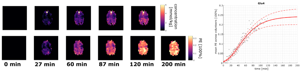
Ziegs T, Ruhm L, Wright A, Henning A. Mapping of glutamate metabolism using 1H FID-MRSI after oral administration of [1-13C] Glc at 9.4 T. Neuroimage 270, 119940 (2023)
2H MRS & MRSI
Deuterium magnetic resonance spectroscopy (2H MRS) is a non-invasive method that allows for the detection of heavy water and deuterated fatty acids based on the 0.015 % natural abundance of the MRI detectable isotope 2H. Furthermore, the enrichment of 2H labeling can be observed in glucose, glutamate and lactate in the brain and body after intake of 2H labeled glucose. Respective enrichment time courses obtained by time resolved experiments enable the calculation of in vivo metabolic turnover rates including the glycolytic rate and the Krebs cycle rate. 2H MRS can be applied for human as well as preclinical studies.
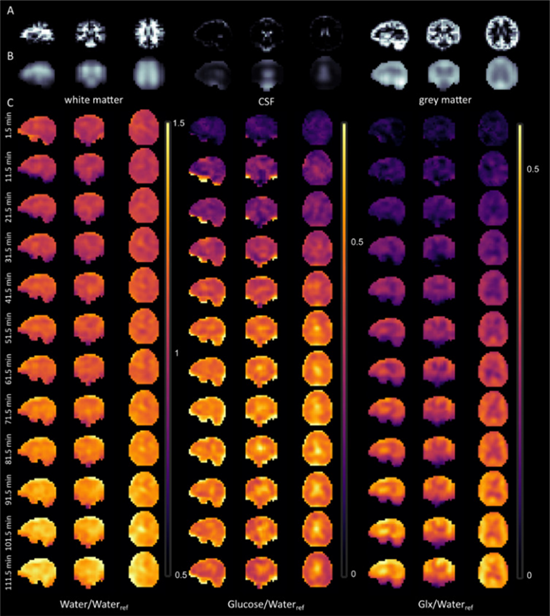
Ruhm L, Avdievich N, Ziegs T, Nagel A, De Feyter H, de Graaf R, Henning A. Deuterium Metabolic Imaging in the human brain at 9.4 Tesla with high spatial and temporal resolution. NeuroImage 244, 118639, pp. 1 - 12 (2021)
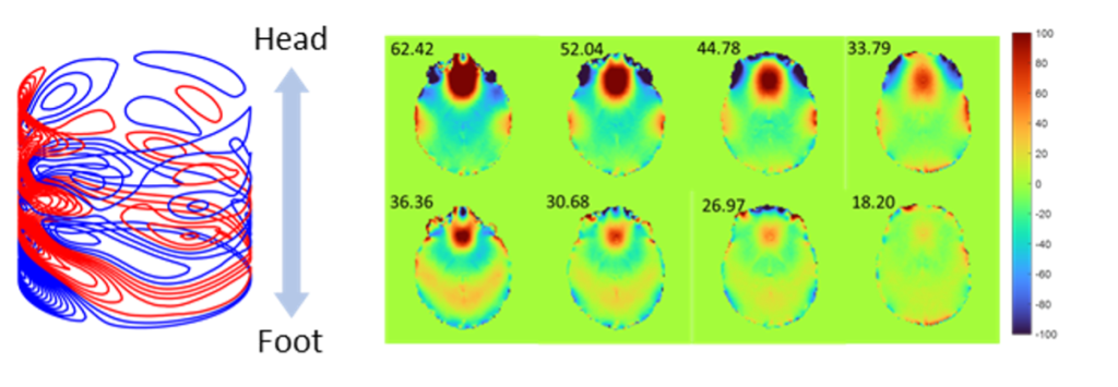
Chemical Exchange Saturation Transfer (CEST)
Chemical Exchange saturation transfer imaging measures the exchange of protons between macromolecules and tissue water. Thus the tissue macromolecular content can be non-invasively determined. A specific focus is the detection of proteins via the amide proton transfer (APT) contrast. In addition CEST imaging allows for measuring intermolecular interactions via the Nuclear Overhauser effect (NOE), chemical exchange rates, metabolite content and pH.
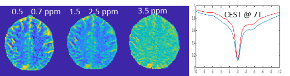
Demetriou E, Dimitrov IE, van Zijl P, Hoogduin H, Vinogradov E, Zhang B, Henning A. Demonstration of B1 mapping based RF shimming for CEST imaging at 7T. Proceedings of the 31st annual scientific meeting of ISMRM, June 3th – 08th 2023, Toronto, Canada.
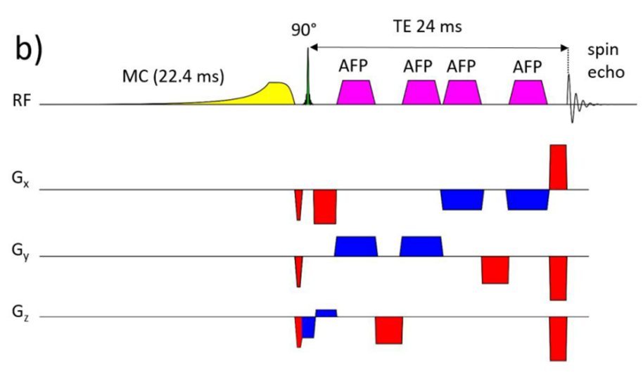
Novel Data Acquisition Methods
MR spectra can be obtained from a single localized volume of interest or metabolic images can be derived from spectroscopic imaging. Robust localization, encoding and artefact suppression methods are developed to ensure optimal data quality. In addition, edited and 2D spectroscopy techniques are implemented to reduce spectral overlap and to increase the number of detectable metabolites. The information encoded into CEST data on the other hand is very sensitive to the applied RF field strength. Hence RF shimming and parallel transmission along with fast encoding schemes and measuring exchange rates are evaluated for CEST. All metabolic MRI methods are intrinsically slow due to the need to encode and resolve the metabolic information paired with a low signal to noise ratio. Hence acceleration of the data acquisition while conserving signal strength is another important research focus and neural network based reconstruction approaches are evaluated.
Dorst, J.; Borbath, T.; Landheer, K.; Avdievich, N.; Henning, A.: Simultaneous detection of metabolite concentration changes, water BOLD signal and pH changes during visual stimulation in the human brain at 9.4T. Journal of Cerebral Blood Flow and Metabolism 42 (6), pp. 1104 - 1119 (2022)
Novel Data Analysis Methods
Preprocessing, image reconstruction and quantification pipelines are of outermost importance for accurate and precise metabolic readouts. Comprehensive single voxel and spectroscopic imaging reconstruction and processing pipelines are developed along with own spectral fitting software and quantification pipelines for proton and non-proton MR spectroscopy. Highlights of these developments are a neural-network based spectroscopic imaging reconstruction method and the development of a spectral fitting package that outperforms the gold standard in the field. In addition, novel spectral models are developed for macromolecules, downfield spectra and labeled isotope data and spatially resolved metabolic modelling is attempted.
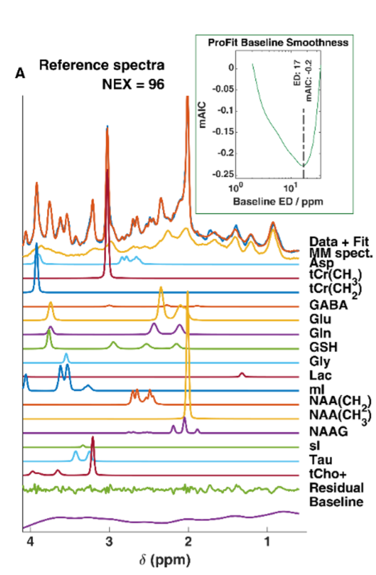
Borbath, T.; Murali-Manohar, S.; Dorst, J.; Wright, A.; Henning, A.: ProFit-1D-A 1D fitting software and open-source validation data sets. Magnetic Resonance in Medicine 86 (6), pp. 2910 - 2929 (2021)
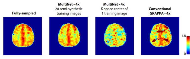
Chan KL, Ziegs T, Henning A. Improved signal-to-noise performance of MultiNet GRAPPA 1H FID MRSI reconstruction with semi-synthetic calibration data. Magnetic Resonance in Medicine 88 (4), pp. 1500 - 1515 (2022)
Ultra-High Field MRI
Increasing the sensitivity and spatial resolution of noninvasive medical imaging methods holds great clinical value potentially providing disease information not available in current clinical practice. Magnetic resonance imaging (MRI) operating at field strength beyond 3 tesla (T), also known as ultrahigh field MRI, generates submillimeter spatial resolution, deciphers cortical layer specific responses of the brain to environmental stimuli and provides previously unavailable metabolite information in a clinically feasible time. The Advanced Imaging Research Center hosts the 7T MRI - the highest clinically approved MRI field, and develops cutting-edge enabling technologies in areas including novel contrast, data acquisition and reconstruction, imaging hardware and calibration methods. We strive for the clinical translation of these promising technologies through collaboration with clinicians on campus and worldwide. While MRI systems with a B0 field strength of 7T and above (i.e. ultra- high field (UHF)) provide substantial advantages and potentially improves diagnostic sensitivity and specificity compared to clinical field strength, various technical challenges of UHF systems have to be overcome before they can be routinely used in the clinic.
Radiofrequency (RF) coil design
NE2.511 is home to a state-of-the-art RF engineering laboratory equipped with full-wave simulation servers, precision circuit routers, dielectric probes, network analyzers, 3D printers, and other specialized equipment for design, prototyping and construction, as well as for diagnostics and repairs of RF coils, anatomical housings, and biomorphic phantoms. Our RF core staff have the expertise and instrumentation to build workhorse clinical coils as well as specialized multinuclear coils and novel designs such as high-impedance ultra-flexible coils. The primary missions of RF lab in AIRC are to: 1) use basic physics research to understand the interaction of RF coils and tissues to improve the sensitivity, transmit efficiency, coverage and safety of RF coils; and 2) develop application-specific coils to facilitate research projects at high and ultra-high field strength. One prime example of our in house RF coil design is a 7T head & neck coil with extended longitudinal coverage as compared to the standard commercial 7T head coil available on the market.

Zhang B, Lowrance D, Henning A. Simultaneous Head and Cervical Spinal Cord Imaging at 7T with a 16-channel transceiver loop array. Proceedings of the 31th annual scientific meeting of ISMRM, June 3th – 08th 2023,Toronto, Canada.
Parallel Transmission
The shorter electromagnetic wavelength at UHF, which results in inhomogeneity of the radiofrequency (RF) field can lead to spatially varying flip angles and, thus, a spatial variation of the image contrast, signal dropouts or brightening. The most flexible approach to overcome this issue is parallel transmission, which enables much improved control over the spatial and temporal RF field by exploiting the additional degrees of freedom of multiple independent RF transmission channels in combination with spatial encoding via gradients. AIRC researchers explore the concept of universal parallel transmit pulse design with optimized gradient trajectories and applicable to a large flip angle range. This in turn will enable the application of this approach to various clinically important MRI sequences.
(Left) Geldschläger O, Bosch D, Henning A. OTUP-workflow: Target specific optimization of the transmit k-space trajectory for flexible universal parallel transmit RF pulse design. NMR in Biomedicine 35 (8) e4728 (2022) (Right) Boga C, Geldschlaeger O, Henning A. Quasi-Damped Newton’s Method for Large Flip Angle Universal Pulse Design Using Parallel Transmission at 9.4T. Proceedings of the 31th annual scientific meeting of ISMRM, June 3th – 08th 2023,Toronto, Canada.
B0 Shimming
B0 field inhomogeneity is one of the most severe problems in ultrahigh field human MRI. Homogeneity of the static magnetic field (B0) is required in order to obtain artefact free images in functional and metabolic MRI scans. Various artifacts can result from poor B0 homogeneity, such as signal dropout, geometric distortion, curved slice profiles and insufficient peak separation in MR spectroscopy. On one hand a versatile stand-alone custom written B0 shimming tool (MATLAB) was implemented. Complementary to this software solution we performed numerical simulations to optimize wire patterns for a dedicated head shim insert. With this hardware brain B0 shimming can be largely improved in comparison to using the scanner integrated 2nd order spherical harmonics B0 shimming only.
Su Shengyue, Henning A. Design of human brain shim coil with high-density wire pattern translated from Stream Function. Proceedings of the 31th annual scientific meeting of ISMRM, June 3th – 08th 2023,Toronto, Canada
Multimodal High-Field MRI for Clinical Research
Metabolic MRI methods can be used to determine mean tissue concentrations of metabolites and compounds along with pH, ion concentrations and metabolic turnover rates. In addition, the change of all these parameters over time upon disease progression, treatment response, functional tasks, exercise or 13C or 2H labeled isotope intake to measure metabolic rates can be evaluated.
Hence metabolic MRI methods are used for clinical diagnostics as well as neuroscientific, physiological and clinical trials. They are generally applicable to the human brain, different organs in the body including spinal cord, liver, kidney, breast, prostate and heart as well as the skeletal muscle and adipose tissue.
Emerging clinical applications of ultra-high field MRI include the detection of small lesions and structures in epilepsy, multiple sclerosis and cartilage degeneration and mostly utilize high resolution anatomical imaging. The high spatial resolution and contrast of functional magnetic resonance imaging at ultra-high fields enables the visualization of complex inter- and even intra-cortical networks. Ultra-high field MRI also allows for highly spatially resolved metabolic imaging and the detection of a much enlarged number of metabolites and compounds. Multimodal MRI at >7T is thus highly valuable to study psychiatricm neurological and neurodegenerative disorders, brain cancer, breast cancer, prostate and gynecological cancer, spinal cord disease, liver metabolism, joints as well as inborn genetic defects.
Brain and Spinal Cord Imaging
Proton and phosphorous MR spectroscopy and spectroscopic imaging provide rich information about the integrity of neuronal and glial cells, neurotransmission, brain energy metabolism, cell membrane turnover, protein content and antioxidant levels. These techniques are widely used to study psychiatric, neurological and neurodegenerative disorders, traumatic injury of the brain and spinal cord, inborn genetic defects in children as well as brain cancer. The examples shown below are: (I) anatomical and 1H spectroscopic imaging data of brain cancer; (II) single voxel 1H MRS data demonstrating metabolic changes in spinal cord injury; (III) changes in neurotransmission in mild cognitive impairment and (IV) change in energy metabolism in major depression.
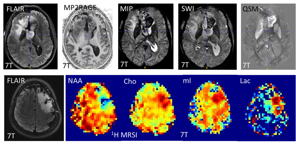
(top) Anatomical MRI of brain cancer patient shows abnormal blood vessel growth. (bottom) Metabolic MRI using 1H MRSI in brain cancer patient shows reduced N-acetyl-aspartate and elevated choline, myo-inositol and lactate. Data were acquired in the context of CPRIT funded brain cancer trial at 7T.
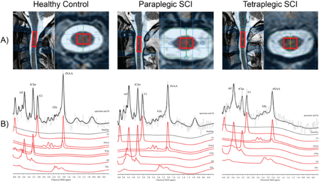
Wyss PO, Huber E, Curt A, Kollias S, Freund P, Henning A. MR Spectroscopy of the Cervical Spinal Cord in Chronic Spinal Cord Injury. Radiology. 2019 Apr;291(1):131-138.
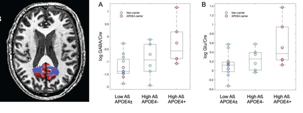
Simon J. Schreiner, Thomas Kirchner, Jiri M. G. Van Bergen, Stefanie C. Steininger, Michael Wyss, Anton F. Gietl, Alfred Buck, Valerie Treyer, Roger M. Nitsch, Christoph Hock, Klaas P. Pruessmann, Anke Henning, Paul G. Unschuld. Gray matter GABA and glutamate indicate genetic, pathological, and clinical risk of Alzheimer disease.
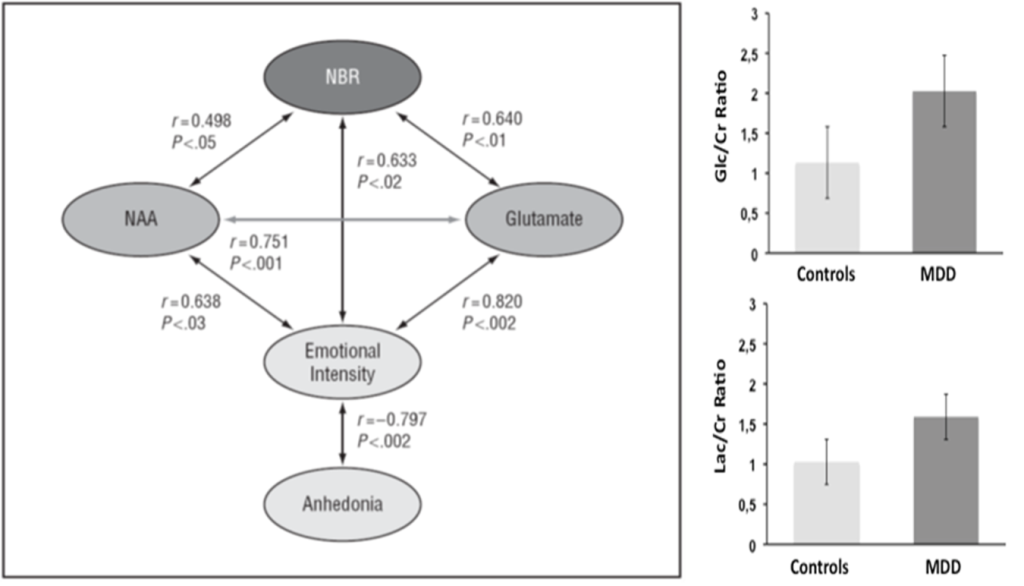
(left) Walter M*, Henning A*,Grimm S*, Schulte RF, Beck J, Dydak U, Schnepf B, Boeker H, Boesiger P, Northoff G. The Relationship Between Aberrant Neuronal Activation in the Pregenual Anterior Cingulate, Altered Glutamatergic Metabolism, and Anhedonia in Major Depression. Archives of General Psychiatry 66(5):478- 486, 2009. (right) Ernst J, Hock A, Henning A, Seifritz E, Boeker H, Grimm S. Increased pregenual anterior cingulate glucose and lactate concentrations in major depressive disorder. Molecular Psychiatry: 22(1) 113–119, 2017.
Cardiac and muscle 1H and 31P MRS
MR spectroscopy in the myocardium can reveal changes in energy metabolism and energy storage. AIRC researchers have expertise with respect to scan protocol development for 1H and 31P single voxel MRS in the human myocardium that include optimal static magnetic field shimming, artefact suppression and frequency stabilization. Previous clinical trials using 31P MRS revealed changes in the myocardial energy metabolism in acute and subacute Tako-Tsubo cardiomyopathy, hypertrophic cardiomyopathy, diabetes mellitus type 1 as well as heart failure with preserved ejection fraction and partial reversal of the effect by respective treatments. It was also shown for the first time that the discrimination of lipid in the heart muscle cells from lipid of adipose tissue surrounding the heart is possible by 1H MRS in the human myocardium, which opens the possibility to investigate the impact of energy storage in the metabolic syndrome.
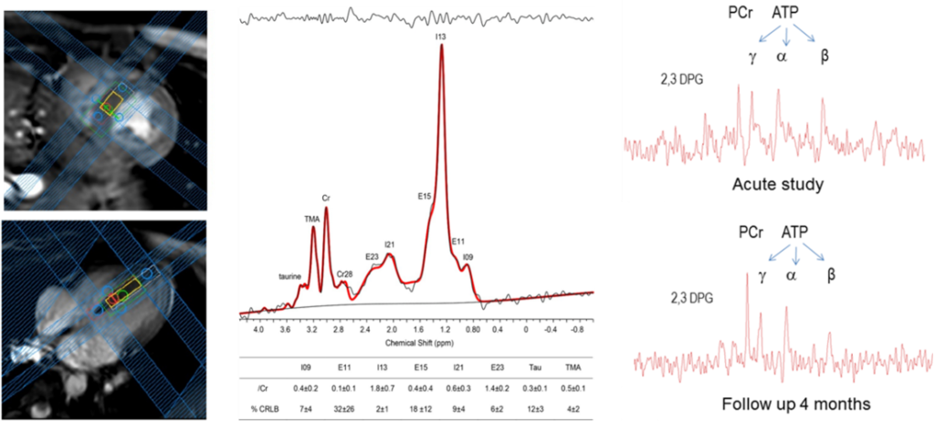
(left) Fillmer A, Hock A, Cameron D, Henning A. Non-Water-Suppressed 1H MR Spectroscopy with Orientational Prior Knowledge shows potential for separating of Intra- and Extramyocellular Lipid Signals in Human Myocardium. Scientific Reports 7(16898) 1-14 2017. (right) Scully C, Rudd A, Mezincescu A, Wilson H, Srinivasan J, Horgan G, Broadhurst P, Newby D, Henning A, Dawson D. Persistent Long-Term Structural, Functional, and Metabolic Changes After Stress-Induced (Takotsubo) Cardiomyopathy. Circulation 137 (10), pp. 1039 - 1048 (2018)