Research
Our research projects include developing innovative hydrogel biomaterials, and applications of these biomaterials to understand and modulate cell-matrix interaction, improve ocular, nueral and liver tissue engineering and regeneration


The interplay between cell and micro-nano features can be leveraged to control a range of cell functions. During my PhD and postdoctoral research work, I made significant contributions in understanding the effect of patterned biomaterial topography on the functions of vascular endothelial cells, smooth muscle cells and neuronal cells. I studied the differential response of vascular endothelial and smooth muscle cells on nano-patterned TiO2 – a widely used vascular-stent material. I found that smooth muscle proliferation can be suppressed by patterning the TiO2 with 70 nm gratings, which can potentially reduce the restenosis rate. In another collaborative study, I investigated the effect of aligned nano-topographies on the migration rate of diabetic and healthy endothelial cells.
High-resolution patterning of topography on hydrogel surface is highly challenging. To achieve this, I developed a new hydrogel nano-imprinting process, which made it possible to ask: how does the topographies on soft-tissue-like stiffness induce morphological changes in cells? Using my patterned hydrogel, the team discovered that the topographies cause focal-adhesion polarization, which is critical to induce cellular alignment on aligned topographies. Lastly, during my postdoc at University of Waterloo, my mentored MS student (S. Mattiassi) and I identified topographies that improved the maturation of neurons obtained from direct conversion of fibroblasts.
The human pluripotent stem cell-derived hepatoblasts, when co-cultured with Jagged1-expressing (Notch ligand) mouse cells, differentiate into liver bile duct cells. Yet, the co-culture with mouse cells diminishes the translational potential of human cells, and does not allow spatio-temporal control of Notch pathway, which is required to construct complex liver models. To replace the need for mouse cells, I developed a Jagged1-immobilized hyaluronan hydrogel that successfully differentiated hPSC-derived hepatoblasts into bile ducts cells. I discovered that the biological function of full-length Notch ligands (Jagged1, DLL1) can be masked by attaching streptavidin photo-cages to the ligands via a photo-cleavable linker, which was a conceptual leap. This enabled the spatio-temporal control of Notch signaling by using timed photo-exposure in different regions of cell culture, paving the way to develop complex cell-culture models requiring advanced control of Notch signaling. This work was done in collaboration with another talented chemist postdoc, Dr. Fokina, who optimized the synthesis of photocleavable linker.

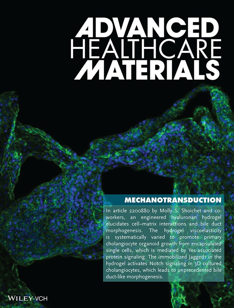
At the University of Toronto, I developed a defined hyaluronan-laminin hydrogel for 3D culture that enabled the growth of liver bile-duct organoids from single cells isolated from mouse bile ducts. I uncovered the stress-relaxation rate of the hydrogel, which was varied by varying the amount of laminin in the hydrogel, drastically affected the growth of single cells into bile-duct organoids. I demonstrated that the faster stress-relaxing microenvironment activated the Yes-associated protein (YAP), which was critical for liver bile duct organoid formation. Inhibition of YAP significantly abrogated the organoid formation. Moreover, I found that the FAK-Src complex was also involved in the mechanosensing of the 3D microenvironment. Lastly, I demonstrated that the Notch signaling and the fast-relaxing microenvironment work synergistically to initiate branching and tubular bile duct formation: Notch signaling alone, or fast-relaxing microenvironment alone, did not induce tubular bile duct formation. Overall, these findings provided the first mechanistic insight into the growth of liver bile duct organoids in a well-defined 3D hydrogel.
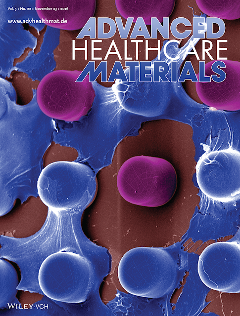
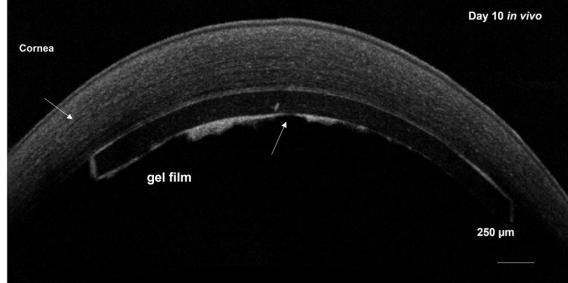
Around 40% of corneal transplantations are performed due to failure of the corneal endothelium alone. The primary human corneal endothelial cell (hCECs), which are isolated from donor corneas, are critical to develop tissue-engineering strategies for corneal-endothelium patients. However, hCECs are extremely difficult to cultivate in vitro because of the loss of functional phenotype. My PhD work directly addressed the overlooked role of cell-surface interactions in the proliferation of primary hCECs in vitro and their transplantation. I discovered that the substrates patterned with micro-nano topographies induced ~3X faster proliferation in donor hCECs, while maintaining their functional characteristics. To transplant the hCEC monolayers, I developed a new nano-patterned hydrogel that supported the growth of the hCEC-monolayer. The hydrogel was made exceptionally stronger using a new sequential-crosslinking approach of gelatin. Due to high strength, the hydrogel could be fabricated in the form of a thin film (critical for corneal transplantation), and could be transplanted into rabbit eyes through minimally-invasive surgery without damaging the hCEC-monolayer. Lastly, to understand the effectiveness of a cell-injection therapy approach for corneal endothelial patients, I created the first in-vitro model of Fuchs Dystrophy (corneal endothelial disease). I achieved this by analyzing the microstructural changes on the diseased corneal surfaces and recapitulated this surface by using innovative microfabrication approaches. This in-vitro model enabled the systematic investigation of simulated cell-injection approaches in patients with different stages of the disease. Using this model, I determined that the cell-injection therapy is likely to be least effective in patients with advanced stage of the disease.

