Current & Ongoing Projects
Hormonal and Mechanical Contributions to Pregnancy-Induced Remodeling of Pelvic Floor Muscles
Funding: NIH K99/R00 HD115224, NIH T32 HD007203
Vaginal childbirth is a significant risk factor for the later development of pelvic floor disorders, though years to decades separate these events. In order to understand what happens to the pelvic floor muscles (PFMs) during vaginal childbirth, we must first understand their structure and function at the onset of delivery. This is especially important as studies have shown that pregnancy protects soft tissues from being injured by large tensile strains. Thus, we're investigating how hormones and mechanical loads contribute to pregnancy-induced remodeling of these skeletal muscles.
In one arm of this study, we isolated muscle stem cells (MuSCs) and fibro-adipogenic progentors (FAPs) from PF and tibialis anterior (TA, hind limb) muscles taken from non-pregnant, mid-pregnant, and late-pregnant female Sprague-Dawley rats and allowed them to differentiate in culture. In addition to looking at differences between groups, we also had conditions where cells were exposed to isolated and combined hormones (progesterone, estrogen, testosterone) to see which may be contributing to any pregnancy-induced differences.
In another arm, we harvested whole muscle samples from the same pregnancy stages and performed ex vivo active and passive mechanical testing. After, samples were flash frozen in OCT for later immunohistochemical assessment. From this data, we can evaluate pregnancy-induced changes in active and passive PF and TA muscle behavior and correlate those differences with variables representing tissue structure and composition (e.g., average muscle fiber cross-sectional area and collagen content).
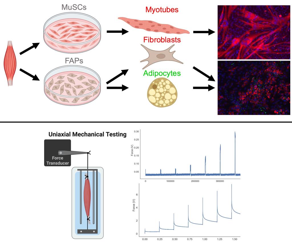
Top) An illustration depicting the cell culture methods used in this study: isolation of muscle stem cells (MuSCs) and fibro-adipogenic progenitors (FAPs), differentiation in culture, and immunohistochemistry for quantification. Bottom) An illustration depicting a whole muscle uniaxial mechanical testing setup and representative graphs of the types active and passive mechanical data collected.
Bupivacaine-Induced Regeneration of Extraocular Muscles
Funding: NIH T32 HD007203, Strabismus Research Foundation
Strabismus involves misalignment of the eyes, and treatments often focuses on the extraocular muscles (EOMs) - the group of individual muscles that surround the eye and control eye movement. Bupivacaine injection is a non-surgical treatment option that is generally effective, though the mechanisms of its effects on the EOMs remain unclear. Thus, we are collaborating with Dr. Jolene Rudell to investigate the impact of bupivacaine injections on EOM structure and mechanical function.
Thus far, we've found that, initially, bupivacaine has a myodestructive effect on 3-month-old New Zealand white rabbit EOMs injected with 3% bupivacaine solution compared to saline and non-injected controls. However, at 1-week post-injection, the bupivacaine-injected EOMs demonstrate more embryonic myosin heavy chain positive fibers, indicative of muscle regeneration; and at 3-months, this group demonstrates a greater density of muscle stem cells and more myofibers with centralized nuclei compared to saline controls. These findings suggest that bupivacaine triggers a regenerative response in injected rabbit EOMs, contributing to knowledge describing how bupivacaine impacts EOMs to treat strabismus.
Read about Bupivacaine-Induced Regeneration in Rabbit Extraocular Muscles: Implications for the Treatment of Strabismus published in Ophthalmology Science in 2026.
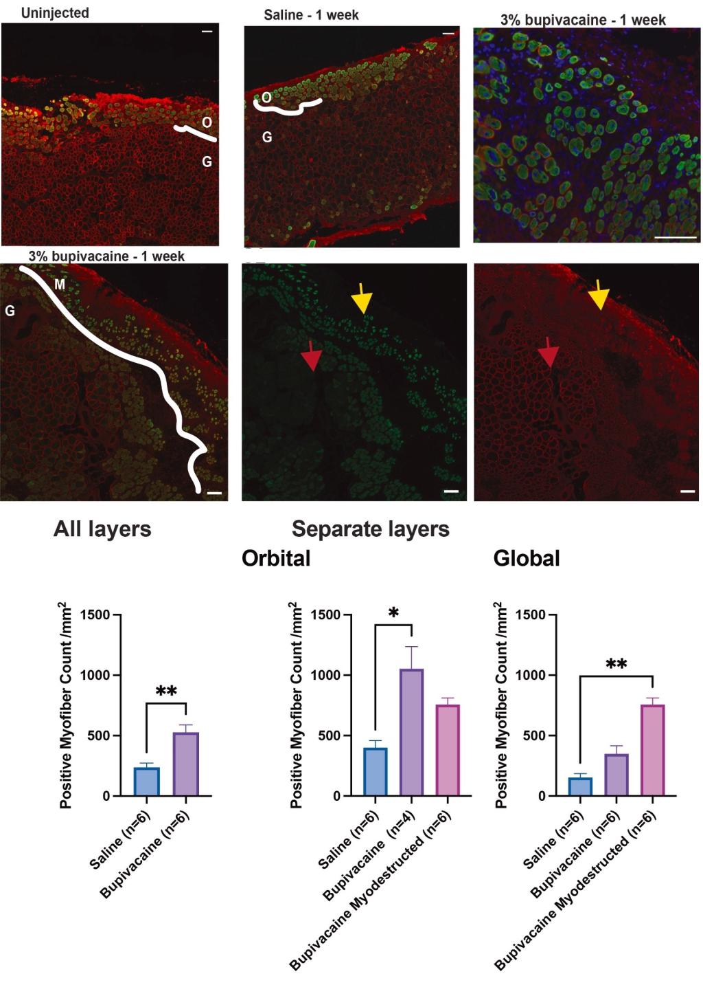
Top) Representative immunofluorescence images of extraocular muscles (EOMs) showing dystrophin (red, outlines the myofiber sarcolemma) and embryonic myosin heavy chain (green, fills newly regenerated myofibers). Bottom) Plots visualizing the number of myofibers per area positive for embryonic myosin heavy chain and demonstrating increased numbers with bupivacaine injection.
Past Projects
Select Studies from Dr. Routzong's Postdoctoral Research at UCSD
Postdoc Advisor: Dr. Marianna Alperin - University of California, San Diego
Sex-Based Differences in Rat and Human Pelvic Floor Muscle Architecture
Funding: NIH T32 HD007203
The Sprague-Dawley rat is an established animal model for studying the female pelvic floor muscles (PFMs), however, its suitability for the study of male pelvic floor muscles and sex differences is undetermined. Thus, we aimed to determine 1) whether pelvic floor muscle architecture exhibits sexual dimorphism in rats and/or humans, and 2) if the male rat is also a suitable animal model for the study of male human pelvic floor muscles. We found that both rat and human pelvic floor muscles exhibit sexual dimorphism and determined that the Sprague-Dawley rat is also a suitable animal model for the study of male pelvic floor muscle architecture.
Read about Sexual Dimorphism in the Architectural Design of Rat and Human Pelvic Floor Muscles published in the Journal of Biomechanical Engineering in 2024.
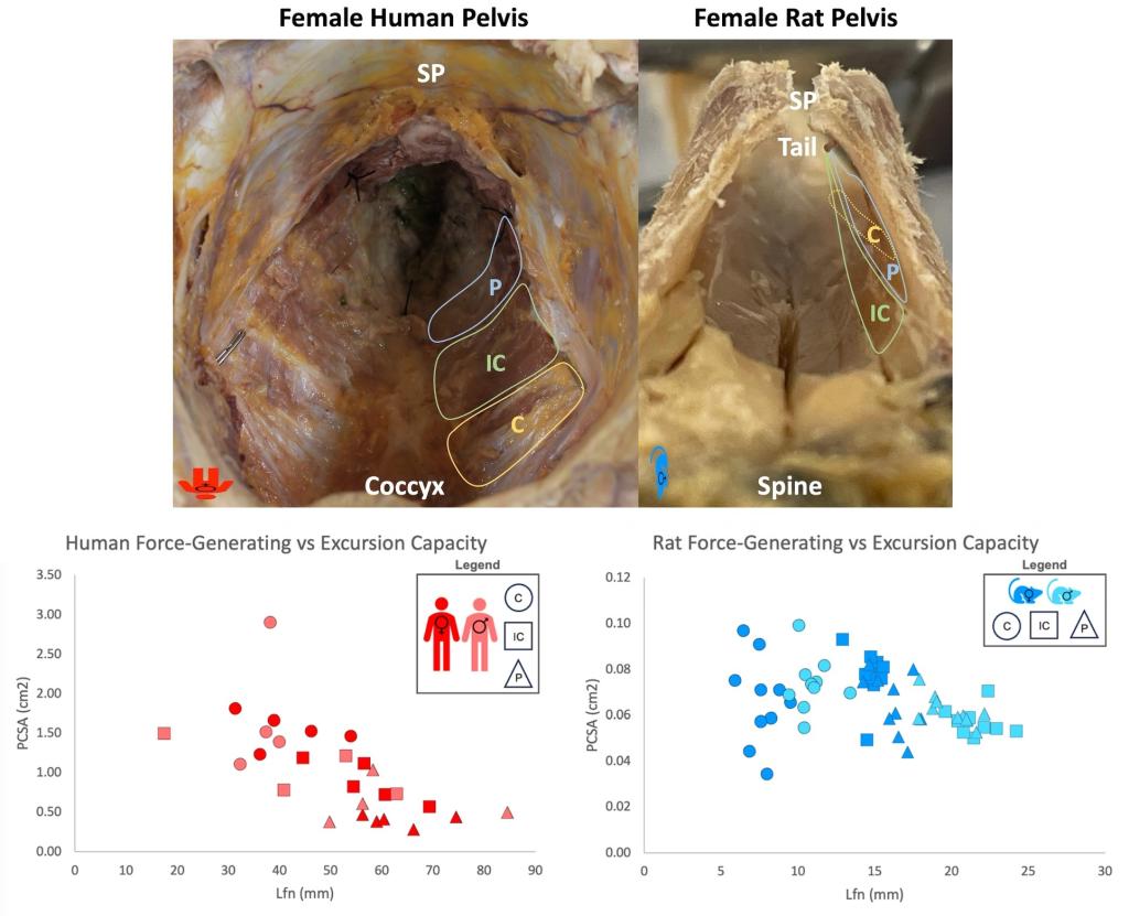
Top) Top down views of a fixed female human (left) and female rat (middle) pelvis with the PFMs exposed, demonstrating general differences in anatomy between species. Bottom) Plots of the force-generating vs excursion capacity for each muscle group, a relationship calculated based on skeletal muscle architecture but indicative of in vivo function.
Select Studies from Dr. Routzong's Doctoral Research at Pitt
PhD Advisor: Dr. Steven Abramowitch - University of Pittsburgh
You can find additional details and studies in Megan Routzong's doctoral dissertation.
Quantifying the Impact of Superficial Perineal Muscles on Finite Element Simulations of Vaginal Delivery
Funding: NSF GRFP 1747452
Finite element models are useful tools for studying the biomechanics of maternal pelvic floor muscles during vaginal childbirth. However, previously, simulations predominantly focused or exclusively included the levator ani muscles. Thus, the goal of this study was to determine the impact of superficial perineal muscles and connective tissues on finite element simulations of the maternal pelvic floor during vaginal delivery.
We directly compared two nearly identical simulations, with the only difference between them being the inclusion/omission of the superficial perineal muscles and connective tissues. We simulated passage of an ellipsoid (representing a fetal head) through the maternal bony pelvis and pelvic floor muscles. We found that when these structures are excluded, representing the majority of simulations at that time, the model will overestimate pubovisceral muscle enthesis and underestimate perineal body strains. Therefore, we concluded that it is important to include superficial muscles and connective tissues in simulations of vaginal childbirth when maternal pelvic floor muscle strains are outputs of interest.
Read about Novel simulations to determine the impact of superficial perineal structures on vaginal delivery published in Interface Focus in 2019.
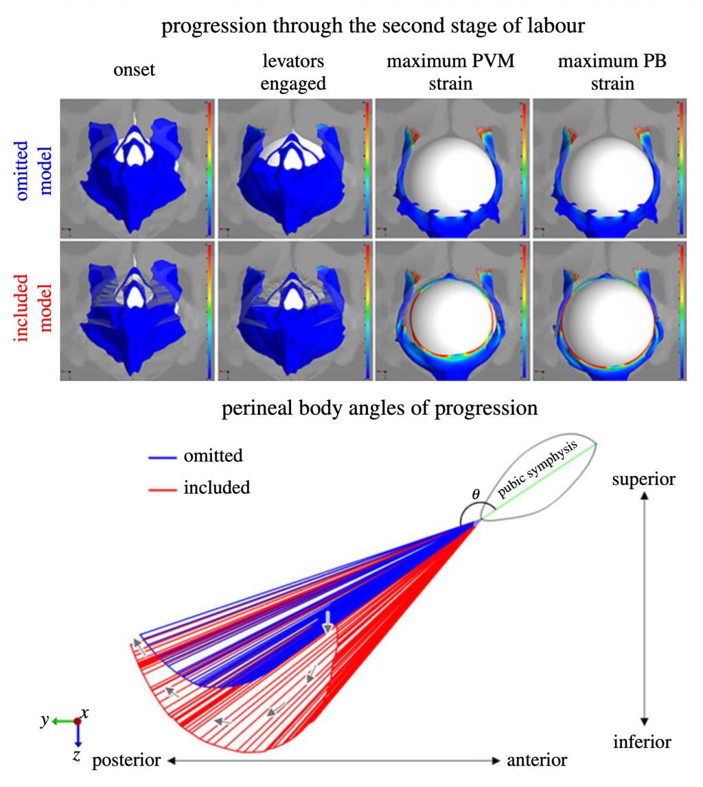
Top) Images depicting pelvic floor muscle strains in each model during simulated vaginal delivery, highlighting larger pubovisceral muscle strains in the omitted model and larger perineal body strains in the included model. Bottom) A visual representation of the perineal body displacements and angles of progression during those same simulations, the motion contributing to larger strains in the included model.
Characterizing the Passive Biomechanical Behavior of Levator Ani and Superficial Perineal Muscles
Funding: NSF GRFP 1747452, NIH T32 HD007203, NIH K99 HD115224
Similar to the above motivation, while there were very few ex vivo mechanical testing studies characterizing female human levator ani muscles (the largest component of the pelvic floor muscles), there weren't any describing the superficial perineal muscles. Thus, we aimed to quantify the passive mechanical behavior of female human superficial perineal muscles via ex vivo mechanical testing and compare it to that of the levator ani muscles.
We obtained levator ani muscle and superficial perineal muscle samples from female human donors and grouped them by muscle type: levator ani, bulbocavernosus, ischiocavernosus, and transverse perineal muscles. We performed uniaxial extension-to-failure tests and used digital image correlation (DIC) to quantify axial and transverse strains. We found that the levator ani muscles exhibit behavior that is distinct from that of the superficial perineal muscles, concluding that levator ani mechanical testing data should not be used to approximate or simulate the biomechanics of superficial perineal muscles.
Read about Passive Mechanical Testing of Female Human Levator Ani and Superficial Perineal Muscles published in Annals of Biomedical Engineering in 2026.
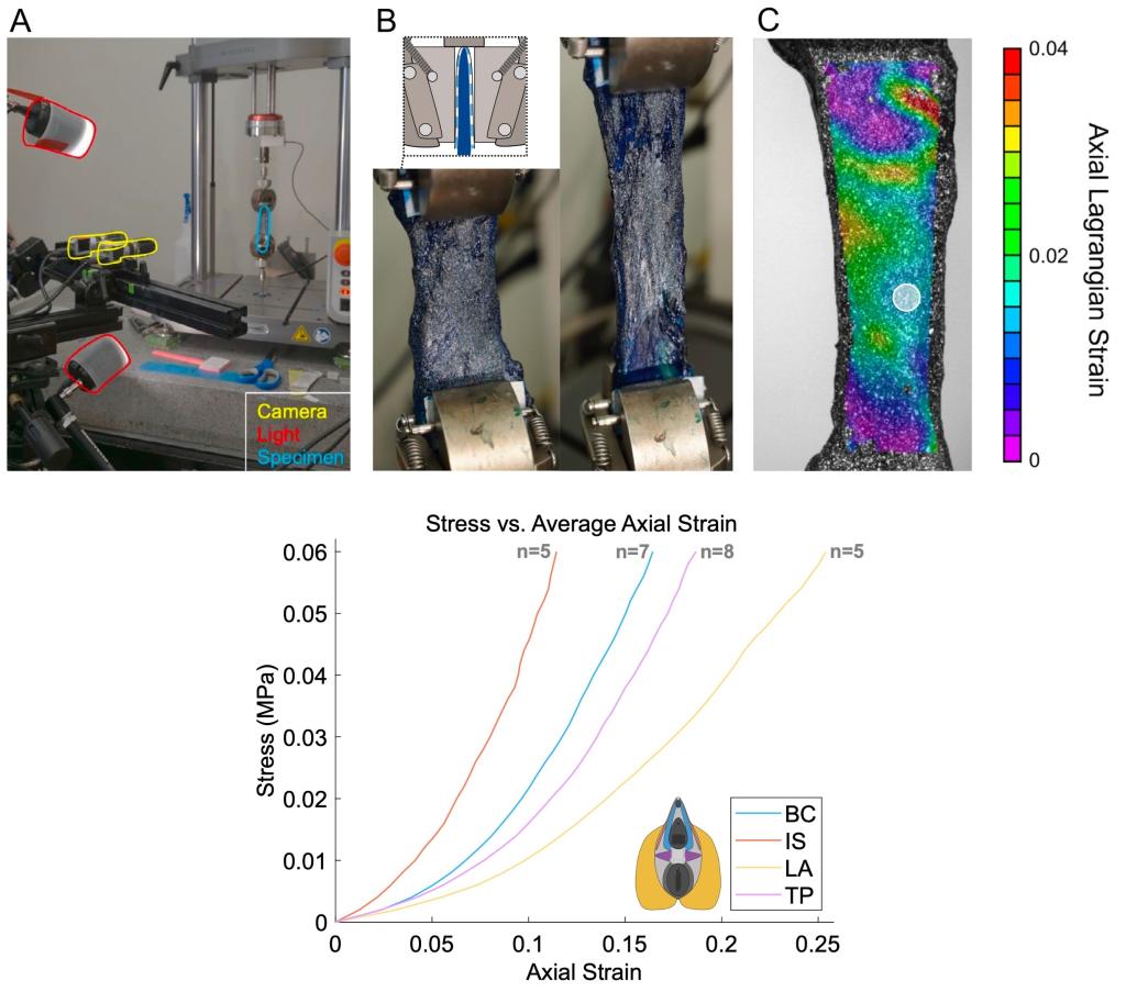
Top) Images and illustrations depicting the mechanical testing and DIC-based strain calculation methods used in this study. Bottom) The average axial stress vs strain curve for each muscle group tested, demonstrating that the levator ani (LA) exhibited the most compliant and the ischiocavernosus (IS) the stiffest response, on average.
Evaluating Female Human Pelvic Floor and Superficial Perineal Muscle Shape Variations via Statistical Shape Modeling
Funding: NSF GRFP 1747452
When assessing biomechanics, shape plays an important role in understanding tissue structure-function relationships. While it was known that pregnancy induces some degree of pelvic floor muscle remodeling, the effect of this remodeling on pelvic floor muscle shape had yet to be robustly explored. Additionally, as pelvic floor muscle shape can be assessed non-invasively, we wanted to explore shape as a metric for the degree of remodeling undergone during gestation. Thus, the goal of these studies was to evaluate midsagittal 2D and 3D pelvic floor muscle shape variations and identify differences between nulliparous (have never given birth), pregnant, and vaginally parous (have given birth vaginally) women using statistical shape modeling.
In the 2D analysis, pelvic floor muscle structures, including the levator plate, anal sphincter complex, and level III vaginal support (i.e., perineal body and superficial perineal muscles/perineal membrane) were outlined in the midsagittal plane. Corresponding points were established and then statistical shape analyses were performed. Early-pregnant (1st and 2nd trimester) shapes significantly differed from late-pregnant (3rd trimester) shapes. Thus, only late-pregnant shapes were included in the final comparison. Meanwhile, shapes from the vaginal parity = 1 group (meaning that woman had one vaginal birth) did not significantly differ from the vaginal parity = 2-4 group, so all were included in the final vaginally parous group. Multiple aspects of pelvic floor muscle shape significantly differed between nulliparous and late-pregnant women. Anatomically, these shape modes described striaghter and more posterior levator plates and anterior protrusion of level III support in late-pregnant compared to nulliparous women while the parous group straddled the other two.
Read about Pelvic floor shape variations during pregnancy and after vaginal delivery published in Computer Methods and Programs in Biomedicine in 2020.
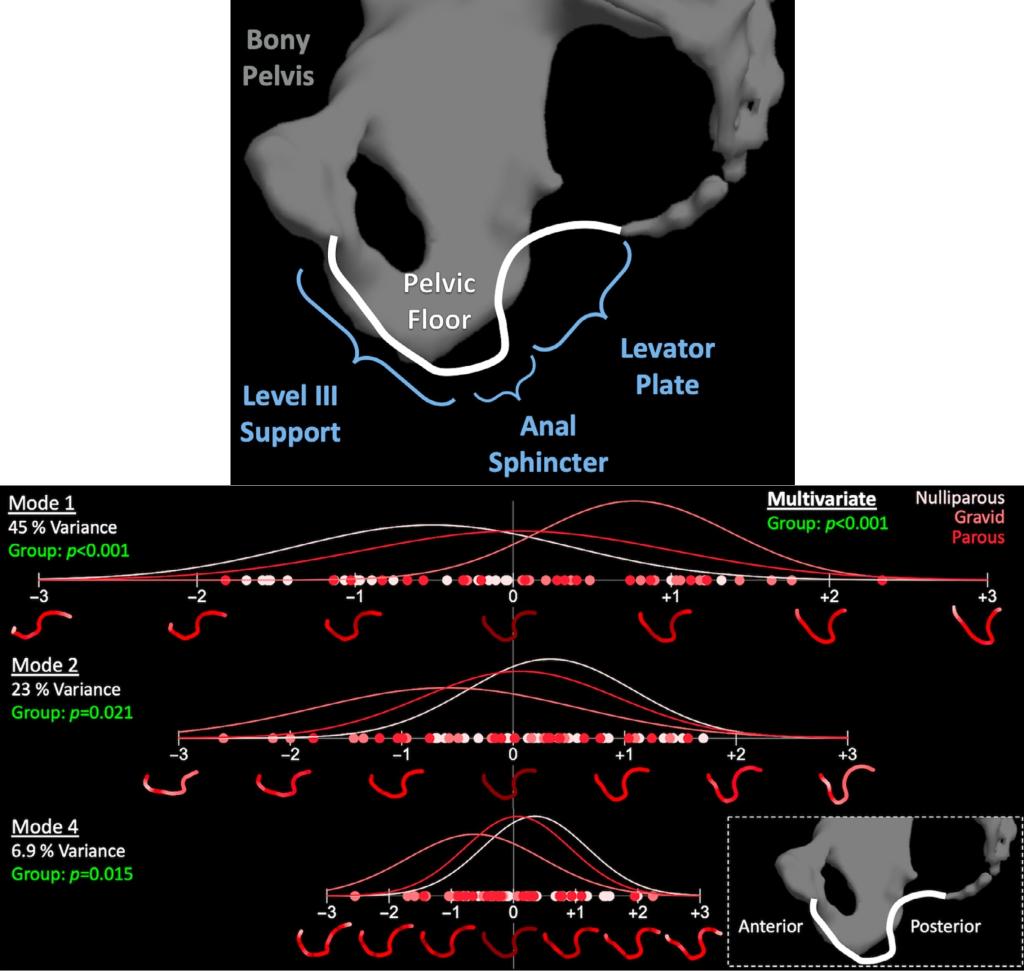
Top) An illustration depicting a 2D pelvic floor muscle polyline with the most relevant anatomy labelled. Bottom) Statistical shape modeling results visualizing nulliparous, late-pregnant (gravid), and parous group distributions across modes 1, 2, and 4 and highlighting how distinct the late-pregnant group generally was from the other groups.
In the 3D analysis, entire pelvic floor muscle complexes, which included skeletal muscle and connective tissue structures, were segmented from retrospectively acquired pelvic magnetic resonance images (MRIs). Corresponding points were established by fitting a high resolution template to each patient-specific shape. Then the same statistical shape modeling workflow as that in the above study was carried out to evaluate population variation and differences between nulliparous, late-pregnant, and parous women. Pelvic floor muscle complex shape significantly different between the late pregnant group compared to the nulliparous and parous groups. This statistical shape analysis described greater perineal and external anal sphincter descent, increased iliococcygeus concavity, and a proportionally wider mid-posterior levator hiatus in late pregnant group, suggesting these tissues/regions are important to consider when evaluating pregnancy-induced changes in pelvic floor muscle structure and function.
Read about Morphological Variation in the Pelvic Floor Muscle Complex of Nulliparous, Pregnant, and Parous Women published in Annals of Biomedical Engineering in 2023.
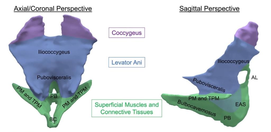
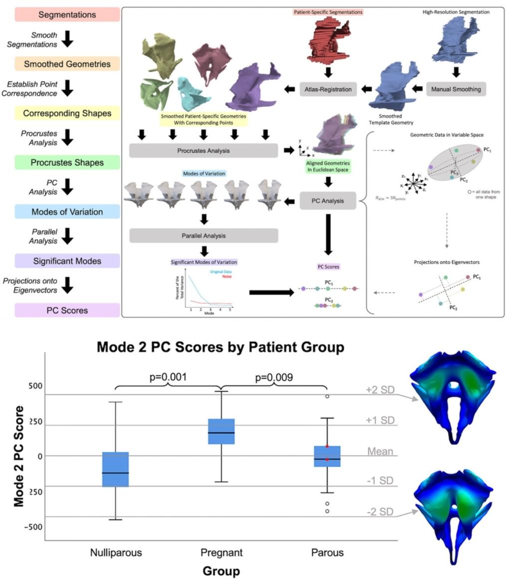
Left) The high resolution pelvic floor muscle template shape with relevant tissues/regions labelled. Top) An illustration of our statistical shape modeling workflow from template creation and segmentation to principal component (PC) score calculation. Bottom) Results for one shape mode depicting PC scores and statistical differences between the late-pregnant compared to the nulliparous and parous groups, and corresponding model outputs demonstrating the physical interpretation of those PC scores (iliococcygeus concavity).