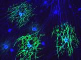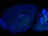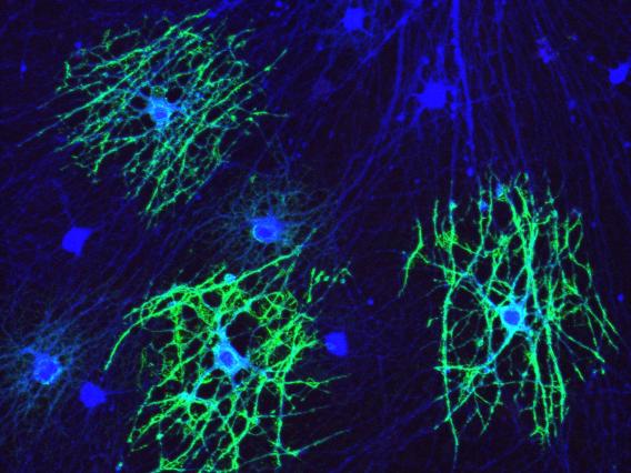
“Myelination on dish” by Lu Sun
Oligodendrocytes (green) co-cultured with retinal ganglion cells (RGC, blue) extended multiple processes to ensheath RGC axons.
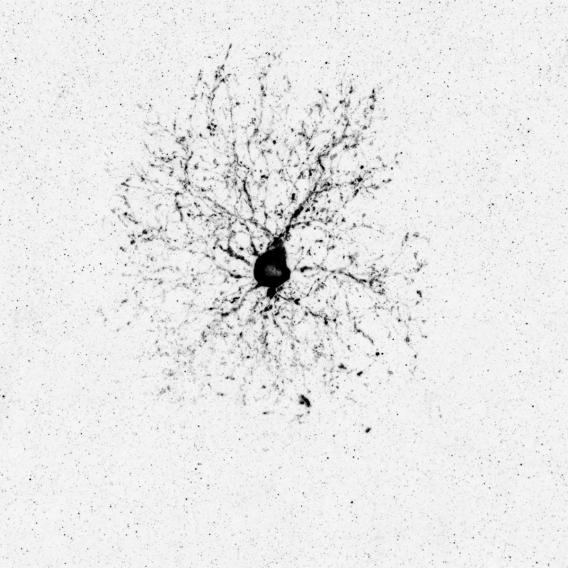
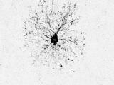
“The intermediate stage during oligodendrogenesis” by Payton Reynolds
A genetic labeled pre-myelinating oligodendrocyte extended numerous processes to possibly search axons for myelination.
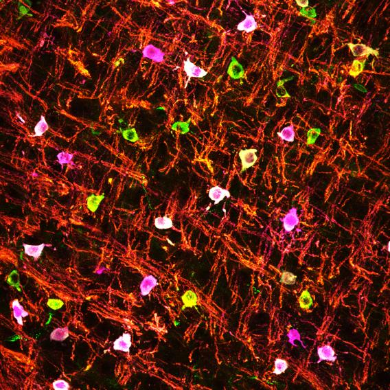
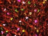
“Myelination in action” by Payton Reynolds
Early postnatal mouse brainstem labeled by MBP (red), CC1 (green), and DsRed (genetically delineated oligodendrocytes in magenta).
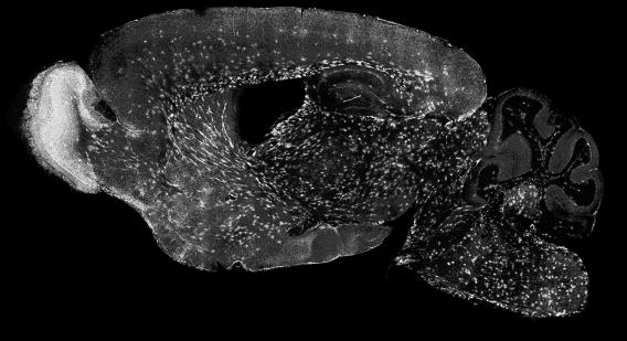
“Tracing myelination in space and time” by Aksheev Bhambri
Subsets of newly generated pre-myelinating oligodendrocytes were genetically labeled and traced for 10 days, showing the spatiotemporal map of oligodendrogenesis at the peak of myelination.
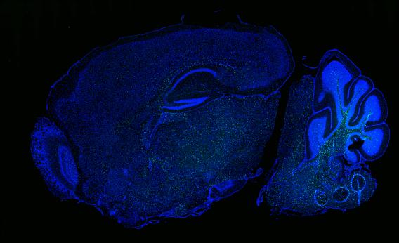
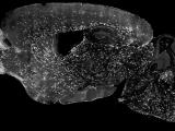
“Visualizing newborn oligodendrocytes” by Aksheev Bhambri
Newborn oligodendrocytes (green, LncOL1) in postnatal day 15 mouse brain section.
