Deep Learning-based Attenuation Correction (DLAC)
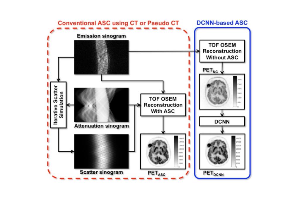
A dedicated brain PET scanner does not come with a combined CT or transmission source, there is no direct solution for accurate attenuation and scatter correction, both of which are critical for quantitative PET. To address this problem, we propose joint attenuation and scatter correction (ASC) in image space for non-corrected PET (PETNC) using deep convolutional neural networks (DCNNs). This approach is a one-step process, distinct from conventional methods that rely on generating attenuation maps first that are then applied to iterative scatter simulation in sinogram space.
Publications:
DLAC for SPECT Myocardial Perfusion Imaging (MPI)
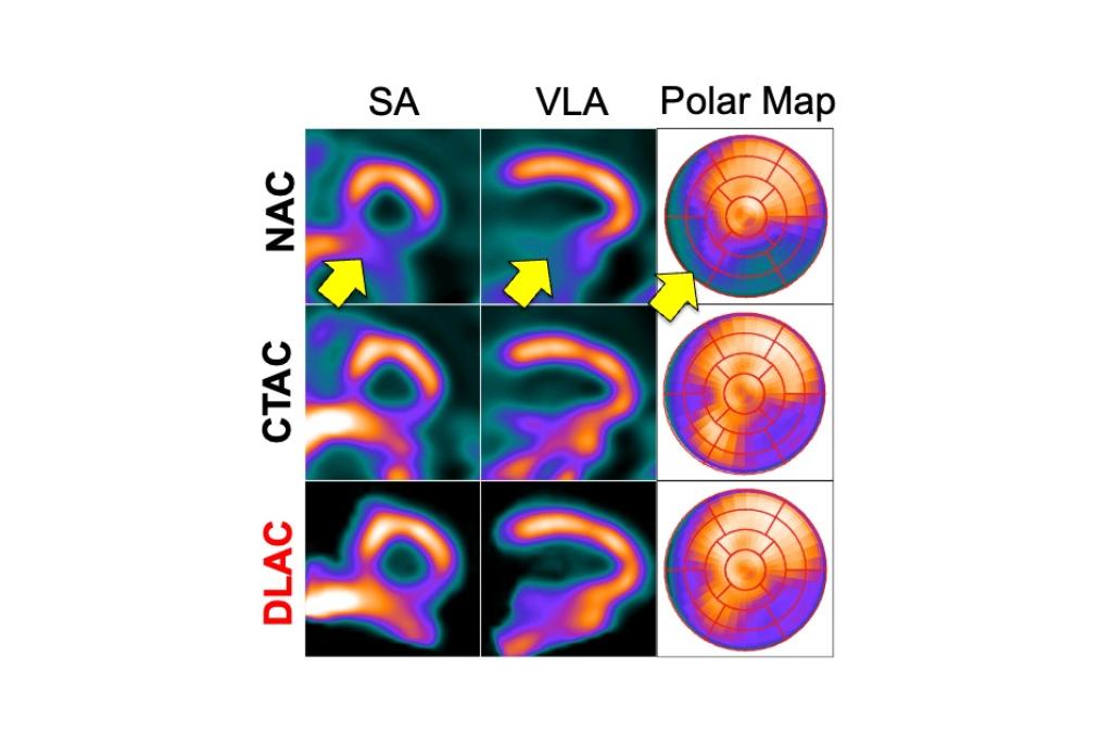
Dedicated cardiac SPECT scanners with cadmium-zinc-telluride cameras have shown capabilities for shortened scan times or reduced radiation doses, as well as improved image quality. Since most dedicated scanners do not have integrated CT, image quantication with attenuation correction (AC) is challenging and artifacts are routinely encountered in daily clinical practice. In this work, we demonstrated a direct AC technique using deep learning (DL) for myocardial perfusion imaging (MPI).
Publication:
Data-Driven Gating (DDG) for PET
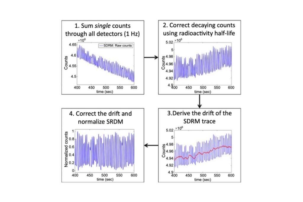
To date, all proposed DDG approaches use coincidences and singles have not been investigated for respiratory motion estimation. In general, randoms and scatters in singles are considered as noisy and needless data to reduce the quality of PET image reconstruction. However, respiratory motion could also be intrinsic to singles such as coincidence events and time-series curves of crystal detectors show signal fluctuation according to respiratory motion. The study demonstrated the feasibility of fast respiratory motion estimation using singles data available as a sorted format in list-mode files acquired in an integrated PET/MRI system for a proof-of-concept.
Publication:
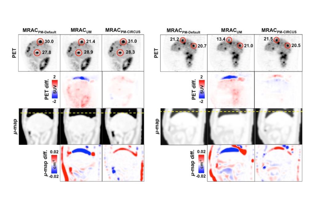
Phase-Matched Attenuation Correction
Respiratory motion causes misalignments between PET and MR-derived attenuation maps (µ-maps) in addition to artifacts on both PET and MR images in simultaneous PET/MRI for organs such as liver that can experience motion of several centimeters. To address this problem, we developed an efficient MR-based attenuation correction (MRAC) method to generate phase-matched µ-maps for quiescent period PET (PETQ) in abdominal PET/MRI. MRAC data was acquired with CIRcular Cartesian UnderSampling (CIRCUS) sampling during 100 s in free-breathing as an accelerated data acquisition strategy for phase-matched MRAC (MRACPM-CIRCUS).
Publication:
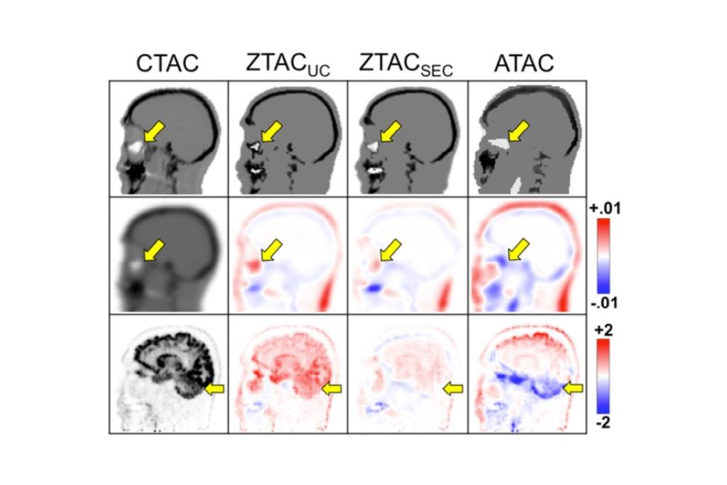
MR-based Attenuation Correction for PET/MR
The ZTE-based attenuation correction (ZTAC) method demonstrated more accurate results in comparison with an atlas-based attenuation correction (ATAC). However, the major challenge of ZTAC was the misclassification of air/tissue/bone mixtures (e.g., sinus or mastoid) or their edges (e.g., skin or trachea) as bone, resulting in overestimated PET quantification. Therefore, it is important to improve the accuracy of ZTAC near the skull base for neurologic PET/MRI studies. Our study evaluated a sinus/edge-corrected (SEC) ZTE-based attenuation correction (ZTACSEC), relative to an uncorrected (UC) ZTAC (ZTACUC) and the ATAC.
Publications:
PET-Guided Tumor Tracking
In this study we evaluated the potential and feasibility of PET for dynamic lung tumor tracking during radiation treatment. The research focus was to evaluate tracking accuracy in a commercial PET scanner for a proof-of-principle based on the assumption of real-time processing. We proposed a center of mass (CoM) lung tumor tracking algorithm using gated-PET images combined with a respiratory monitoring signal. The geometric accuracy of the proposed algorithm was quantified in a dynamic phantom study.
Publication:
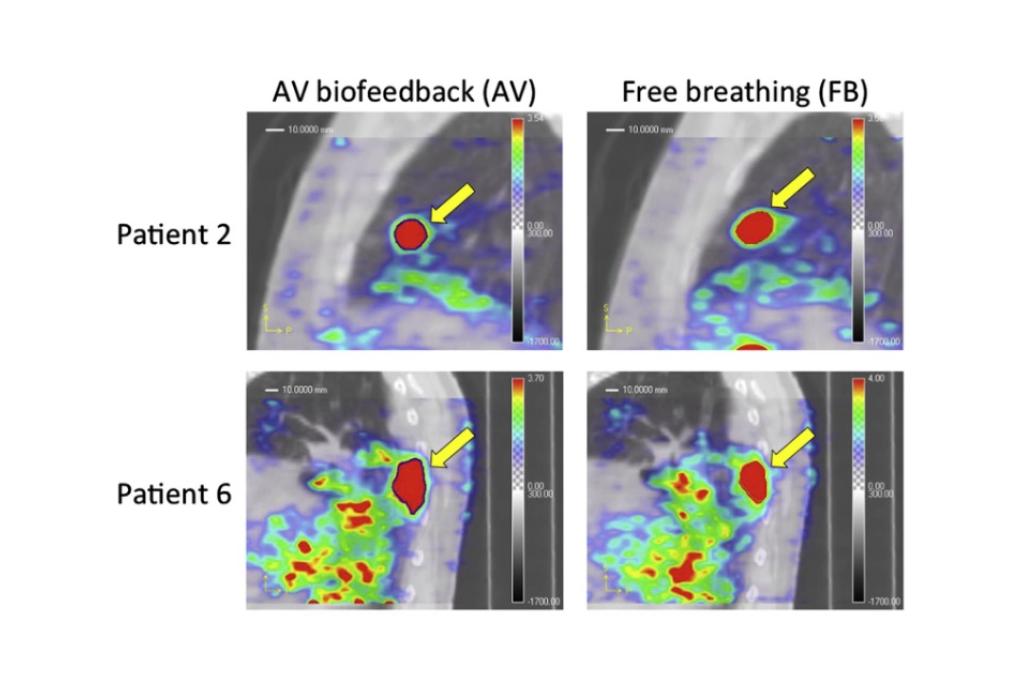
Audiovisual (AV) Biofeedback for Motion Management
Respiratory motion causes artifacts in PET and CT. Thus, respiratory motion management is needed to mitigate such artifacts, which is achieved through four-dimensional (4D) imaging. However, 4D imaging may fail to reduce artifacts if breathing is irregular during data acquisition due to inadequate respiratory motion sampling for image reconstruction. Audiovisual (AV) biofeedback is a real-time, interactive and personalized system designed to help a patient self-regulate their breathing, demonstrating a reduction in the average cycle-to-cycle variations in respiratory amplitude and period. For the first time, the impact of AV biofeedback was evaluated on 4D-PET and 4D-CT imaging in a patient study.
Publications: