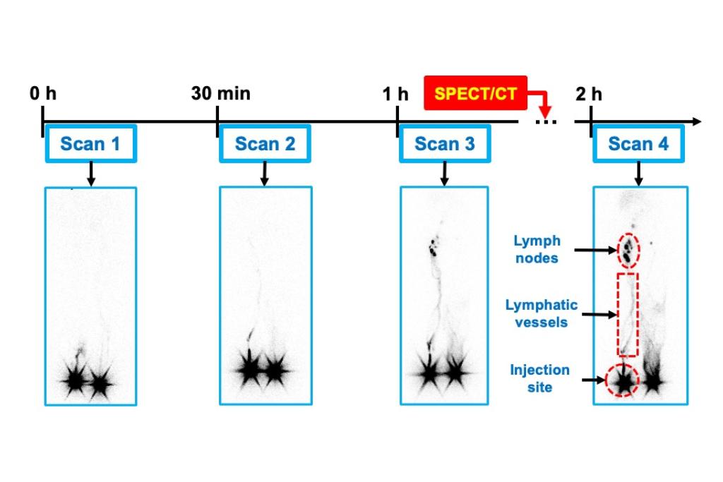
Developing Imaging and Analysis Tools for Lymphoscintigraphy in Lymphedema
Lymphedema occurs when a part of the lymphatic system fails to remove lymph fluid, affecting over 10 million individuals in the United States. Although lymphoscintigraphy (2D planar imaging) has long been regarded as the gold standard with high sensitivity for diagnosing lymphedema, it is limited by noise and lack of quantitative imaging and analysis tools. To overcome these limitations, we are developing a denoising solution along with quantitative imaging and analysis tools to enhance lymphoscintigraphy for lymphedema diagnosis.
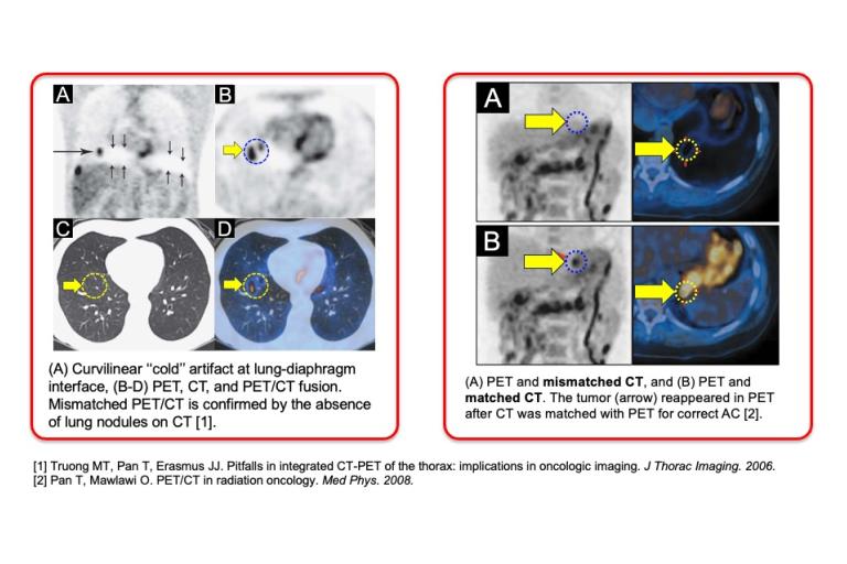
Development of Deep Learning-based Anatomical Matching (DL-AM) for PET/CT
PET/CT is typically performed during free breathing (FB). Because PET acquisition occurs over several minutes, PET images reflect the average anatomical position across many breathing cycles. In contrast, each CT scan captures a snapshot of a specific phase of the respiratory cycle. This mismatch results in visual discrepancies between PET and CT and quantitative bias in PET images. To address frequent anatomical mismatches and respiratory motion artifacts in the chest and abdomen, we propose to develop a DL-based anatomical matching (DL-AM) framework that deforms FB CT images to DDG CT images, achieving accurate alignment with DDG PET.
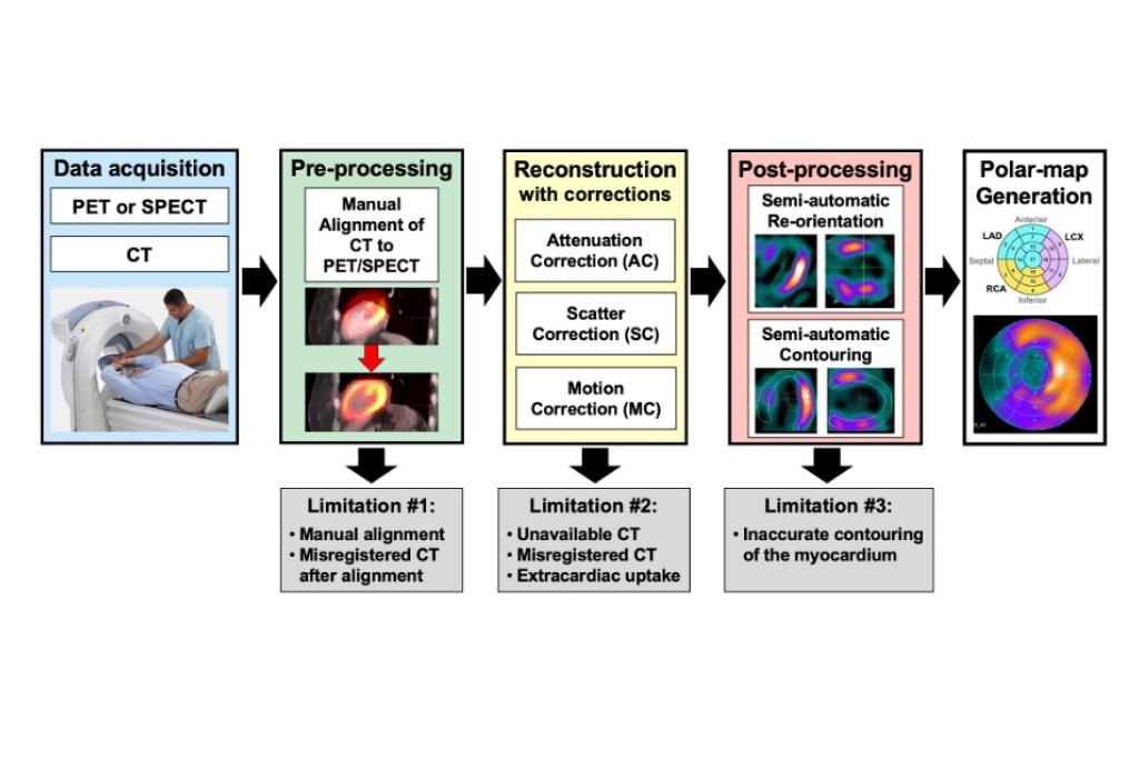
Improving Clinical Workflow in Nuclear Cardiology Using Deep Learning Technology
Nuclear cardiology, using PET and SPECT, is essential for the non-invasive evaluation of cardiovascular disease. However, key limitations persist throughout the workflow, including manual PET/CT or SPECT/CT alignment during preprocessing, inadequate correction methods during reconstruction, and semi-manual myocardial segmentation in post-processing. To overcome these challenges, we propose developing deep learning technologies to enhance each stage—preprocessing, reconstruction, and post-processing—to improve automation, accuracy, and efficiency.