New imaging modalities are necessary for treating deadly diseases
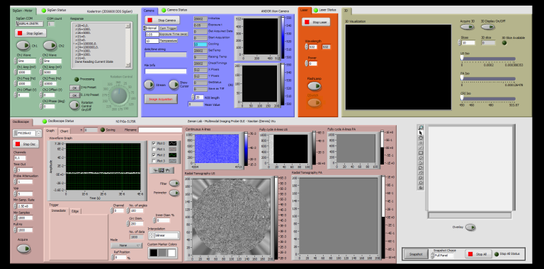
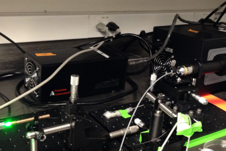
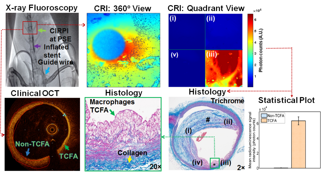
Zaman’s Lab focuses on the design and development of novel cutting-edge multi-mode imaging systems to overcome current limitations in clinical systems. Most recent research project is involved with the design and development of a multimode catheter-based imaging system called a Circumferential Intravascular Radioluminescence Photoacoustic Imaging (CIRPI) for early detection of thin-cap-fibro-atheroma (TCFA), the underlying causes of coronary artery disease, one of the leading causes of morbidity and mortality in the USA and worldwide. Further, the CIRPI system characterizes the plaques based on disease tissue compositions to unravel their complex structures. This CIRPI system integrates optical, photoacoustic, radioluminescence and ultrasound imaging. We seek to better understand the underlying causes of the disease mechanisms. We are dissecting the role of TCFA perturbations on vascular wall processes during atherosclerosis progression. Our lab also studying novel molecular imaging methods to study coronary arterial disease, carotid stenosis, and myocardial ischemia in subcellular level.
 Comparison of different imaging modalities
 Circumferential Intravascular Radioluminescence Photoacoustic Imaging (CIRPI)
The CIRPI system includes a novel optical probe combining circumferential radioluminescence imaging and photoacoustic tomography (PAT). The probe's scintillating imaging window captures radioluminescence images (360° view) of plaques by detecting β-particles during 18F-FDG decay. A tunable laser-based PAT characterizes tissue constituents of plaque at different wavelengths.