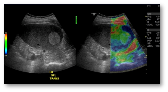
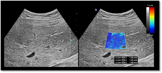
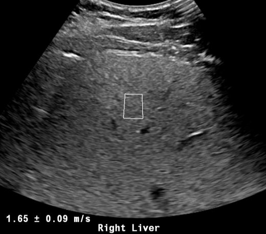
Elastography measures the elastic response of tissues to an externally applied force. Diseased tissues and even cancers are often stiffer than normal tissues. Therefore, elastography may help characterize a tissue by noninvasively measuring its stiffness. It has been used most effectively in liver ultrasound for the detection of fibrosis and is increasingly used to identify cancerous lesions in the breast, prostate, lymph nodes, and thyroid gland.

Shear wave elastography is currently the most advanced method for measuring stiffness. Available on most modern ultrasound machines, shear wave elastography begins by transmitting a high-amplitude “push pulse” (Acoustic Radiation Force Impulse) into the tissue in question. The resultant tissue displacement (micrometer scale) is analyzed by measuring the velocity of shear waves propagating laterally from the ARFI pulse. Tissue stiffness is directly related to shear wave velocity.
Elastography’s benefit lies in its ability to measure tissues that cannot be normally palpated, such as the liver. It also has the advantage of being more uniform across operators and less dependent on operator skill than many other methods of stiffness imaging.

Benefits of elastography include:
- Diagnostically accurate
- Noninvasive; safer than a biopsy
- More cost-effective than many other tests such as CT and MRI
- Can be performed as part of a routine ultrasound study
Ultrasound elastography is FDA-approved for the non-invasive measurement of liver stiffness to evaluate patients with suspected liver disease for fibrosis. Elastography is particularly useful for screening at-risk patients for severe liver fibrosis and cirrhosis, such as patients with hepatitis B and C, and those with hepatic steatosis and non-alcoholic steatohepatitis (NASH). Some medications may also cause liver fibrosis. Faculty at UT Southwestern are also investigating the use of elastography in the differentiation of benign and malignant tumors in the thyroid, lymph nodes, liver, and elsewhere.
