
Dynamin mediated actin bundle

Dynamin on Actin comet tails

Drosophila embryonic musculature

Electron micrograph showing an invasive FCM (pseudo colored light purple) projecting finger-like protrusions into the apposing founder cell.

Wild-type (left) and longer mutant (right) embryos labeled with anti-MHC antibody.

Schematic of Drosophila myoblast fusion. Muscle founder cell attracts fusion competent myoblasts (FCMs) and fuse with them to form multinucleated muscle fibers

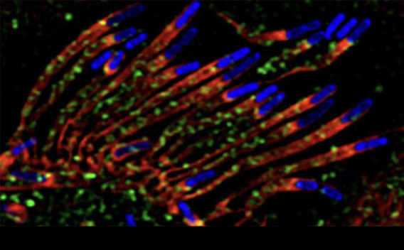
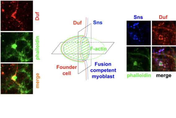
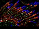
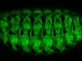
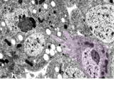
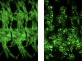
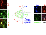
3D representation of an invasive F-actin focus (middle). Horizontal confocal section reveals its invasion into the apposing founder cell (left); and vertical section reveals rings of cell adhesion molecules (Duf and Sns) enclosing the F-actin focus. The F-actin focus and the cell adhesion molecule Sns compose of a podosome-like structure (PLS) at the protruding tip of the FCM.


