Dean et. al., the latest manuscript selected for the cover of Optica. Imaging subcellular dynamics with fast and light-efficient volumetrically parallelized microscopy (Vol. 4, Issue 2, 2017).
Using a combination of structured illumination and TIRF microscopy, the Fiolka lab demonstrated improved optical sectioning in TIRF microscopy (Optics Express, Vol. 24, Issue 26).
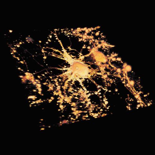
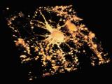
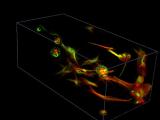
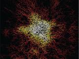
High-speed particle tracking of clathrin-coated vesicles in 3D. Dean, Kevin M., et al., Biophysical journal 110.6 (2016): 1456-1465.
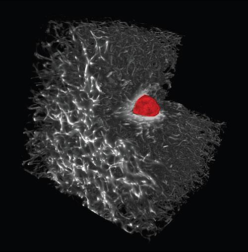
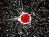
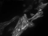
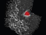
Melanoma cell embedded in collagen.
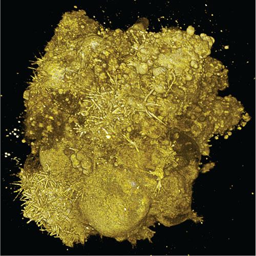
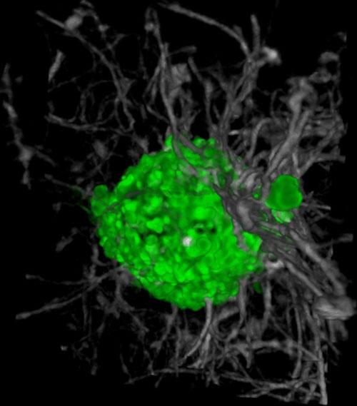
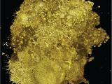
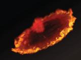
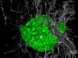
A tumor spheroid consisting of lung cancer cells. Welf, Erik S., et al. Developmental cell 36.4 (2016): 462-475.
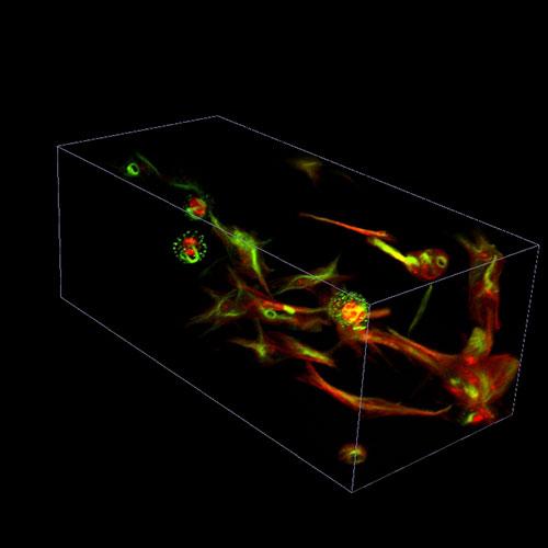
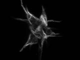
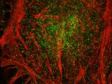
RPE cells, labeled for vimentin and microtubules, embedded in collagen.
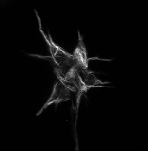
RPE cells, labeled for vimentin, embedded in collagen.
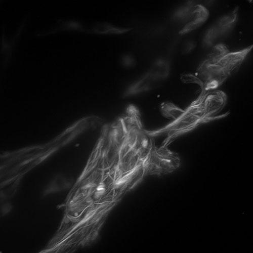
RPE cells embedded in collagen.
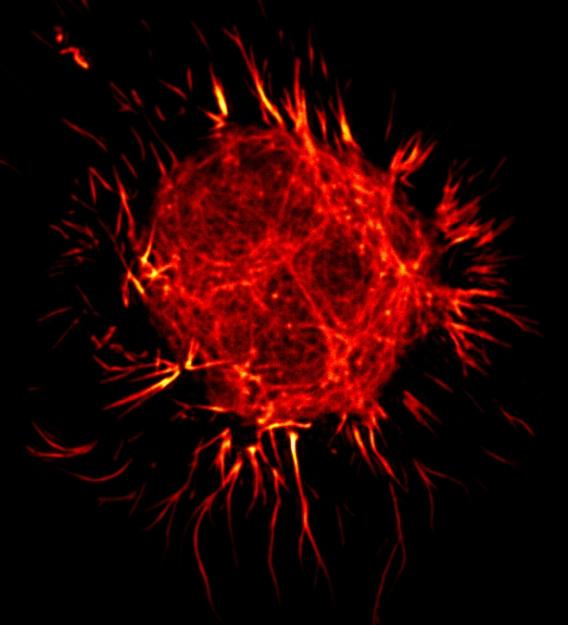
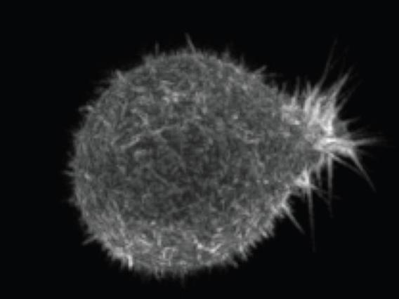
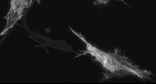
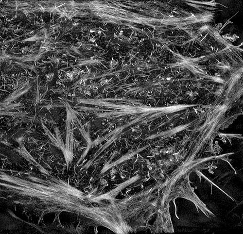
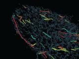
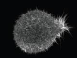
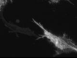
Filipodia in a lung cancer cell. Welf, Erik S., et al. Developmental cell 36.4 (2016): 462-475.
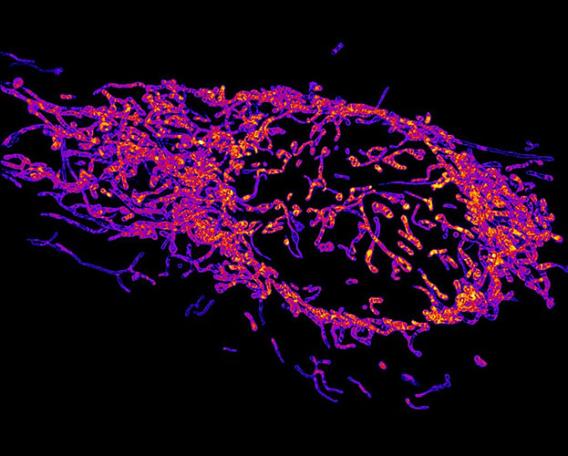
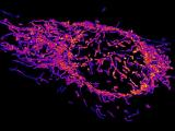
Mitochondria in a HeLa cell imaged with structured illumination.
RPE labeled with tractin-GFP embedded in collagen.
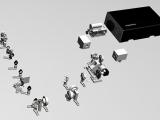
Louis Assembly Render
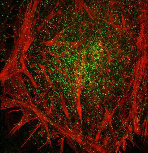
Actin cytoskeleton and clathrin coated pits in a HeLa cell imaged with structured illumination.
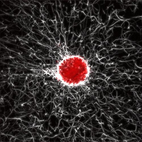
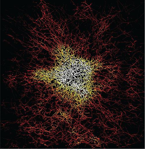
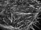
Collagen density surrounding a melanoma cell. Welf, Erik S., etal. Developmental cell 36.4 (2016): 462-475.
3D rendering of melanoma cell in collagen. Welf, Erik S., et al. Developmental cell 36.4 (2016): 462-475.
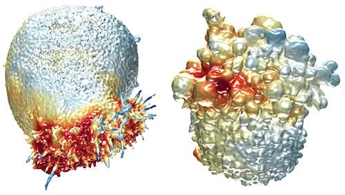
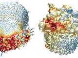
Surface concentration of actin (left) and a biosensor (right) on a lung and melanoma cell, respectively. Welf, Erik S., et al. Developmental cell 36.4 (2016): 462-475.
Rapid volumetric imaging with diagonally scanned light sheet microscopy. Dean, Kevin M., et al. Biophysical Journal 110.6 (2016): 1456-1465.
Actin cytoskeleton in a HeLa cell imaged by structured illumination microscopy.