We study how cells shift from healthy function to early disease by directly visualizing molecular and nanoscale changes inside cells in their native environment.
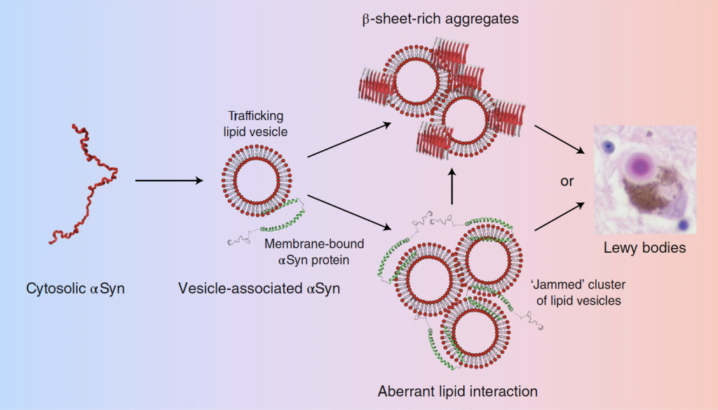
Using cryo-electron tomography of intact neurons and disease-relevant cell models, together with quantitative biophysics and multi-modal imaging, we examine how normally functional proteins—such as alpha-synuclein—misassemble, alter cellular compartments, and destabilize the systems that maintain neuronal health.
To complement these in-cell studies, we also use cryo-electron microscopy to determine higher-resolution structures of disease-associated components extracted from cells, including amyloid fibrils, both alone and in complex with molecular binders selected based on binding affinity.
Our earlier work defined the structure and molecular composition of Lewy bodies and Lewy neurites in human brain tissue. Building on this foundation, we now focus on earlier stages of disease, connecting molecular organization to organelle dysfunction in order to identify the points at which cellular failure first emerges and may still be prevented.
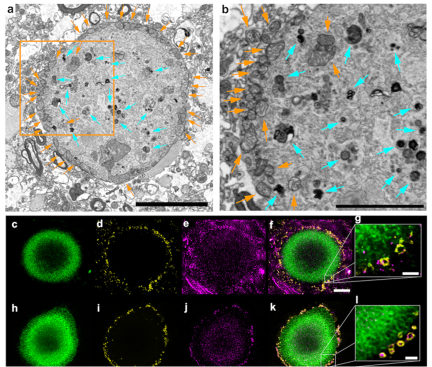 Shahmoradian, Sarah H., et al. "Lewy pathology in Parkinson’s disease consists of crowded organelles and lipid membranes." Nature Neuroscience 22.7 (2019): 1099-1109.