Studying molecular interactions in situ
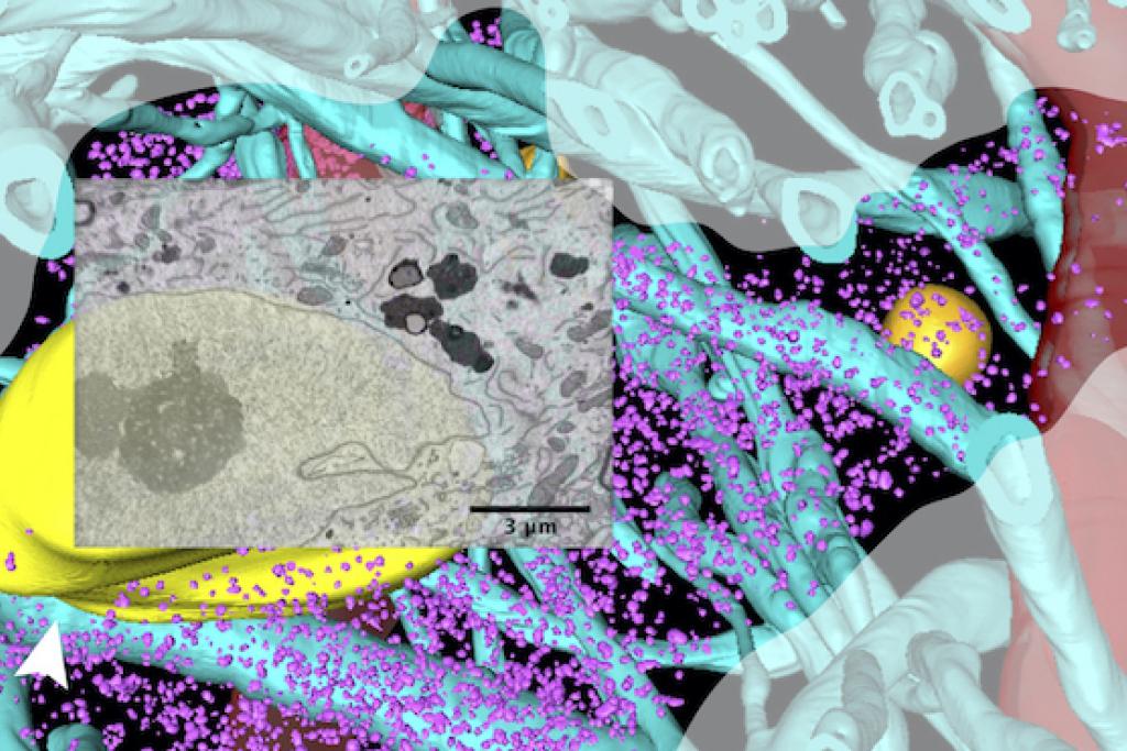 A neuron in human brain tissue surrounded by axons (blue stalks) and blood vessels (red). Overlaid electron microscopy image shows a window into the neuron's internal structure.
 Investigating disease-causing proteins in their native intracellular setting
Parkinson’s and Alzheimer's diseases and other neurodegenerative conditions are hallmarked by the presence of cellular inclusions that arise when proteins misfold and clump into larger, often insoluble structures.
We study the macromolecular basis of how proteins and lipids interact during aggregation: What is their architecture? What precursors do they require? What sub-cellular environment supports them?
What sets our work apart is that we primarily study such aggregation in situ at the nanoscale, which can reveal their subcellular milieu, their components, and how they evolve from the earliest detectable stages, in their natural cellular environment. We primarily use cryogenic-based imaging approaches to this end.
"Seeing is believing", and imaging gives important clues to generate testable hypotheses. To visualize macromolecular complexes both extracted from cells/tissues and within cells/tissues, we use cryo-focused ion beam milling (cryo-FIB) coupled to downstream cryo-electron tomography (cryo-ET) to get nanoscale pictures of cellular processes in a near-native state. Example visualizations here.
We also develop and apply new interfaces and technologies for probing both normal physiology and pathobiology of cellular systems.
We developed a neuronal co-culture cryo-platform uniquely compatible for specialized cryo-fixation that preserves cells in a “glassy” state without chemical fixatives and without ice crystals that destroy tissues and cells. This enables correlative light and electron microscopy (CLEM) of near-native neuronal networks.
These tools, combined with advanced computation, allows to view nanoscale details inside a cell preserved in a near-native state, either healthy or diseased.
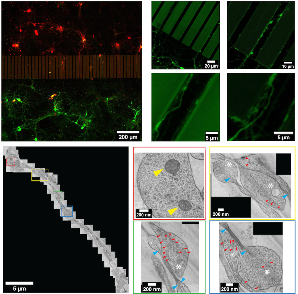 Our microfluidic system for cryo-fixation and CLEM of neuronal networks.
 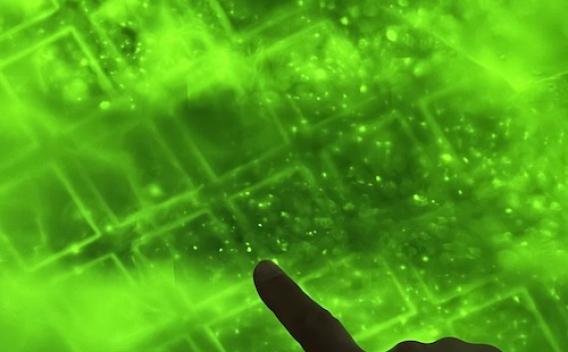 Localizing fluorescently-tagged aggregates within a cellular model of neurodegeneration, using cryo-confocal microscopy.
 Our Research Supporters
We are grateful for the generous research funding from the Parkinson's Foundation, National Institute on Aging (NIA) and the National Institute of Neurological Disorders and Stroke (NINDS) of the National Institutes of Health (NIH), the Lyda Hill Foundation, and the President's Research Council (PRC).