Multi-Layered Valency: Novel Design of Peptidic Imaging Agents for avβ6
NIH R01 EB014244 (MPI: Brown/Sun)
Project goal: To construct multivalent PET imaging probes that target integrin avβ6 for non-small cell lung carcinoma.
Project Description: Synthetic peptides have attracted attention as targeting molecules to deliver radioisotopes to in vivo targets for imaging and/or radiotherapy of diseases. This has been stimulated in part by advances in screening phage-displayed peptide libraries to identify peptidic ligands that target different cell types. Combining advances in peptide ligand development with imaging innovations has the potential to revolutionize the understanding of diseases at the cellular and molecular levels.
Despite the emergence of phage display as a means to isolate cell-binding ligands, a flood of peptide-guided imaging agents has not occurred. This is partially due to the difficulty in retaining the affinity and activity of peptides outside of the context of the phage particle. Moreover, peptides typically have short in vivo half-lives due to degradation and rapid renal filtration2, which limits their accumulation in their targets. Peptide multimerization can improve peptide stability, extend in vivo half-life, and improve affinity. However, most current multimeric peptide constructs are not systematically designed. Aimed to develop a “template” that can be used to rapidly translate peptides isolated by phage display biopanning into molecular imaging probes, this project was proposed to create multivalent peptidic PET imaging agents.
The valency will be modified in three fashions: 1) Multimeric peptide presentation on a common bifunctional chelator scaffold; 2) Multimeric presentation of a peptide sequence on a peptidic construct; and 3) A strategy to combine the multivalent chelator scaffolds with the multivalent peptides to create higher order peptide structures that can be used for molecular imaging.
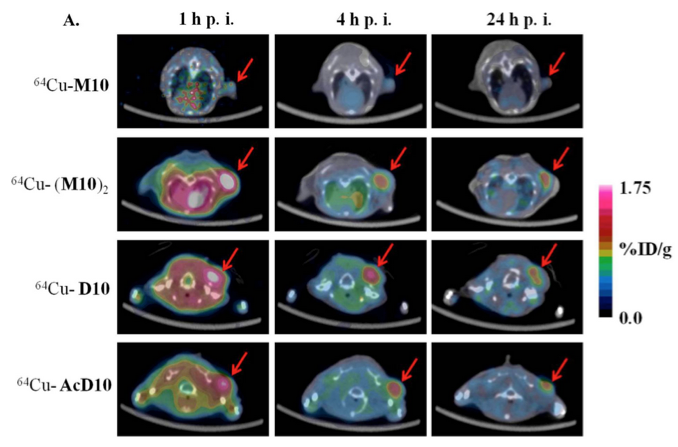
(A) Decay-corrected trans-axial PET images of 64Cu-M10, 64Cu-(M10)2, 64Cu-D10, and 64Cu-AcD10 in SCID mice bearing H2009 tumor at 1 h, 4 h, and 24 h p.i. Tumors are indicated by a red arrow.