Noninvasive Assessment of Pancreatic Beta-Cell Mass.
NIH R01 DK092163 (MPI: Sun/Öz; Key Collaborators: Ahn/Lingvay)
Project goal: To develop a noninvasive imaging technique for clinical assessment of pancreatic beta-cell mass (BCM), a pivotal factor that changes during the progression of diabetes in humans. The ability to noninvasively monitor the fate of BCM under disease and therapy conditions is of particular importance to the prevention and management of diabetes.
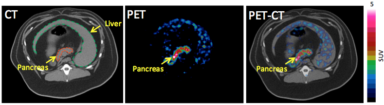
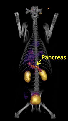
Project Description: Since the first workshop of “Imaging the Pancreatic Beta Cell” co-sponsored by the NIBIB, NIDDK, and JDRFI in 1999, the past decade has seen tremendous progress toward the goal, which is represented by in vivo monitoring of the metabolic fate of islet transplants preloaded with magnetic resonance imaging (MRI) contrast agents and direct imaging of BCM via positron emission tomography (PET). Mainly due to the success of the latter, PET has been regarded with the greatest potential among current imaging techniques to noninvasively monitor the progression of diabetes. However, given the scarcely and heterogeneously distributed beta cell population within the pancreas (1 – 2%) and its gradual mass and function changes in the progression of diabetes, challenges remain in the imaging of BCM regarding reliability, sensitivity, and specificity.
Recently we have successfully developed a bicyclic glucagon-like peptide-1 (GLP-1) analog (EM2198) for specific imaging of BCM when labeled with a positron emitter. To further improve the in vivo behavior and clinical relevance of the probe, multiple copies of the GLP-1 analog are presented on a novel class of bifunctional chelator scaffolds, which consists of a NOTA core (NOTA: 1,4,7-triazacyclononane-1,4,7-triacetic acid) that selectively forms a neutral and kinetically inert complex with 68Ga (t1/2 = 68 min; b+: 90%), a generator-produced isotope.
Noninvasive PET Assessment of Pancreatic Beta-Cell Mass and Function
Juvenile Diabetes Research Foundation 37-2011-632 (PI: Sun; Co-PIs: Öz/Ahn)
Project goal: To develop a noninvasive imaging technique for the assessment of beta-cell function (BCF) in the context of gradual loss of beta-cell mass (BCM) during the type I diabetes progression.
Project description: This project was developed to employ a zinc sensor derivative for specific BCF detection based on glucose-stimulated insulin/zinc co-release in combination with a glucagon-like peptide-1 (GLP-1) based PET probe (EM2198) for specific imaging of BCM. The research strategy is to use a pair of BCM and BCF imaging probes to noninvasively and longitudinally evaluate pancreatic beta-cell function (BCF) in the context of gradual loss of beta-cell mass (BCM) during the type I diabetes progression.