Targeting Tumor Microenvironment for Cancer Imaging and Therapy: Renal Clearable and pH Responsive Gold Nanoparticles.
Cancer Prevention and Research Institute of Texas RP120588 (PI: Zheng; Co-PI: Sun)
Project goal: To design and synthesize a gold nanoparticle-based system for cancer detection and treatment by targeting the tumor microenvironment.
Project Description: Gold nanoparticles (AuNPs) hold great promise in cancer imaging and therapy because of their board material properties and biocompatibility. However, some challenges remain to be addressed for them to be used as anticancer nanomedicine.
This proposal was designed on the hypothesis that through rational design of surface chemistry, particle size, and valence-state gold atom, a new class of IR emitting gold NPs can be created with the following physicochemical properties: 1) highly resistance to nonspecific serum protein adsorption; 2) effective clearance out of the body; 3) pH-dependent adsorption to cell membrane at physiological conditions; 4) in vivo targeting the weak acidic microenvironment of tumors; 5) functioning as a multimodality contrast agent for luminescence, computed tomography and nuclear imaging; and 6) being able to generate singlet oxygen under light excitation to induce the death of cancer cells.
To date, we have shown that a class of 2~5-nm glutathione-coated AuNPs can be reproducibly created with mixed valence states, which exhibited strong triplet-state emission around 580 nm. With little interaction with serum proteins, these AuNPs can be cleared out of the body with an efficiency of 10~100 times better than the same-sized gold nanoparticles. By further modifying the surface chemistry of these luminescent AuNPs, we found that these NPs can bind to cancer cell membranes at pH 6.8 in the presence of serum proteins at the in vitro level while they had no interactions with tumor cells at pH 7.4. Due to strong ligand-metal interactions, these luminescent AuNPs can also photogenerate singlet oxygen with a quantum efficiency of 20%.
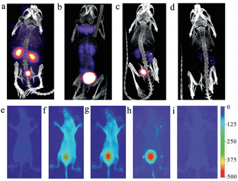
Representative SPECT images (top row) of balb/c mice injected with GS-[198Au]AuNPs. a) 10 min, b) 1 h, c) 4 h, and d) 24 h p.i. In vivo fluorescence imaging (bottom row) of a live mouse e) preinjection, f) 5 min, g) 20 min, h) 1 h, and i) 24 h after IV injection of GS-[198Au]AuNPs.