The long-term goal of our lab's research is to develop and implement imaging technologies that provide unprecedented insight into cancer biology. To increase the realism of cell culture and cancer drug screens, we have abandoned traditional cell cultures on hard glass surfaces and moved towards 3D environments, both ex-vivo and in-vivo. This requires continuous development of dedicated 3D microscope technologies that minimize specimen photodamage and maximize optical penetration depth, image acquisition speed, sensitivity, and spatial resolution.
Clearer View in Microscopy
Optical microscopy is an indispensable tool in the life sciences as it allows minimally invasive observation of biological processes within living cells and organisms with molecular specificity and subcellular detail. In cancer research, optical imaging enables us to dynamically monitor molecular pathways that drive disease, observe cellular responses to pharmacological perturbations, and screen for novel drug effects with high-content readouts of the phenotypic response of cells and tumor tissues over time.
Historically, high-resolution imaging with molecular sensitivity can only be performed under artificial conditions, i.e., cells cultured on glass, even though it has been widely recognized that the cellular microenvironment has a major impact on disease outcomes. In contrast, three-dimensional imaging under physiologically relevant conditions involves stringent trade-offs in spatial and temporal resolution, sample irradiance, and observation time. Further, imaging in physiologically realistic tissues is severely limited by light scattering and sample-induced aberrations.
We aim to extend the current capabilities of optical microscopy by improving the spatiotemporal resolution and optical penetration depth, thereby enabling detailed live cell imaging in physiologically relevant 3D environments. First steps toward this direction have been taken and the results are very promising.
A New Look at Cancer
Cancer research in 3D environments crucially depends on advances in probes, cell culture, microscope technology, and quantitative image analysis. Here at UT Southwestern, we bring all these aspects together via strong collaborations with leading labs in cancer cell biology, microscopy, and quantitative image processing. Through collaborative research, novel microscope technologies developed in my lab are actively used in multiple research programs.
We closely collaborate with other labs to combine our microscope development with their latest quantitative image-analysis tools and cell biological questions. The combination of advanced 3D imaging technology and cutting-edge image processing techniques opens a new window into cancer.
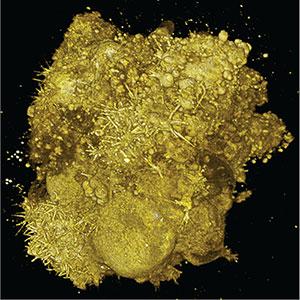 Tumor spheroid consisting of lung cancer cells.