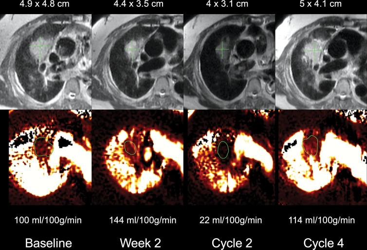
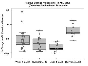
Arterial Spin Labeled Perfusion MRI for the Evaluation of Response to Tyrosine Kinase Inhibition Therapy in Metastatic Renal Cell Carcinoma
We evaluated if arterial spin labeled (ASL) MRI perfusion changes are associated with tumor response and disease progression in metastatic RCC treated with vascular endothelial growth factor receptor (VEGFR) tyrosine kinase inhibitors (TKIs). Seventeen participants received sunitinib (mean age, 59 years ± 7.0 [standard deviation]; 11 men); 11 participants received pazopanib (mean age, 63 years ± 6.6; eight men). Responders had higher baseline tumor perfusion than nonresponders (mean, 404 mL/100 g/min ± 213 vs 199 mL/100 g/min ± 136; P = .02).
Tsai, et al. Radiology 2021
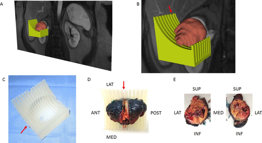
Development of a Patient-specific Tumor Mold Using Magnetic Resonance Imaging and 3-Dimensional Printing Technology for Targeted Tissue Procurement and Radiomics Analysis of Renal Masses
We developed a platform for accurate colocalization of in vivo quantitative multiparametric magnetic resonance imaging features with ex vivo surgical specimens of patients with renal masses using patient-specific 3-dimensional (3D)-printed tumor molds. This approach may aid in targeted tissue procurement and radiomics and radiogenomic analyses.
Dwivedi, et al. Urology 2018
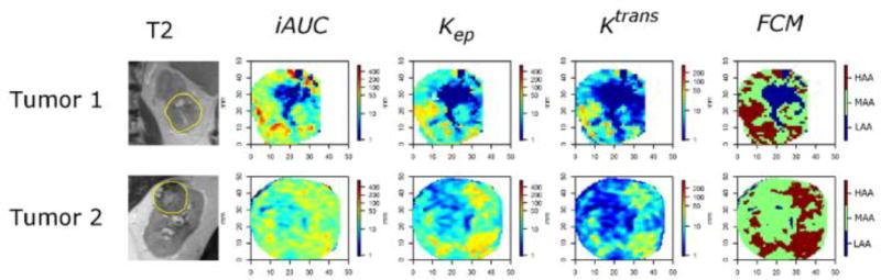
Statistical Clustering of Parametric Maps from Dynamic Contrast Enhanced MRI and an Associated Decision Tree Model for Non-Invasive Tumour Grading of T1b Solid Clear Cell Renal Cell Carcinoma
We developed a novel statistical clustering algorithm, the fuzzy c-means (FCM), to combine information from dynamic contrast-enhanced (DCE) magnetic resonance imaging (MRI) into a single tumor map to distinguish high-grade from low-grade T1b clear cell renal cell carcinoma (ccRCC). A decision tree model using FCM criteria beyond size was developed with 78% accuracy, 86% sensitivity, 73% specificity, 67% positive predictive value and 89% negative predictive value for the detection of high grade in T1b ccRCCs.
Xi, et al. Eur Radiol 2018
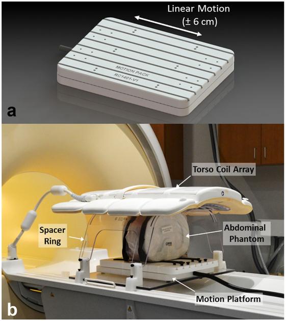
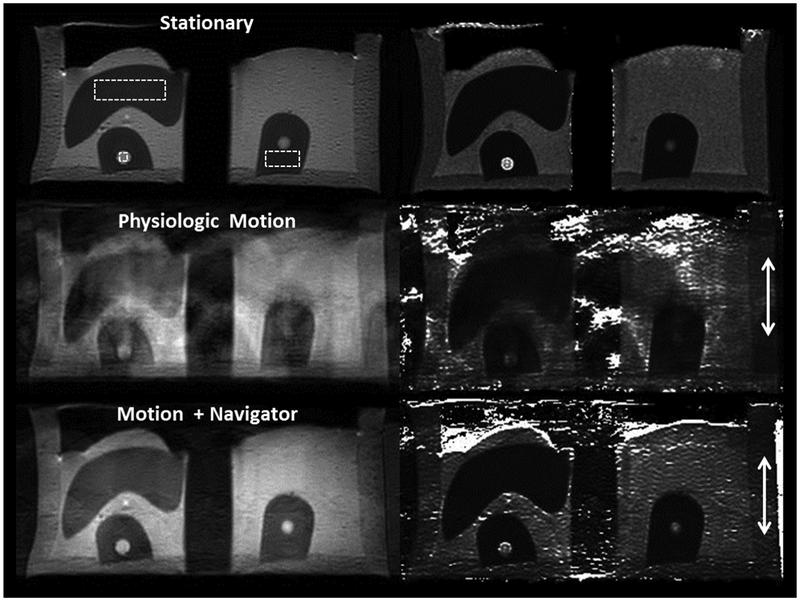
An MRI-Compatible Platform for One-Dimensional Motion Management Studies in MRI
We developed and evaluated a programmable system for one-dimensional motion management in MRI research. The proposed system meets or exceeds the characteristics of motion in the cranial–caudal direction for abdominal organs with respect to displacement, velocity, and acceleration. The system can serve as a foundation for a research platform to investigate and develop motion management approaches for MRI. Subsequent development of this platform has allowed assessment of two-dimensional motion of anthropomorphic phantoms.
Nofiele, et al. Magn Reson Med 2016
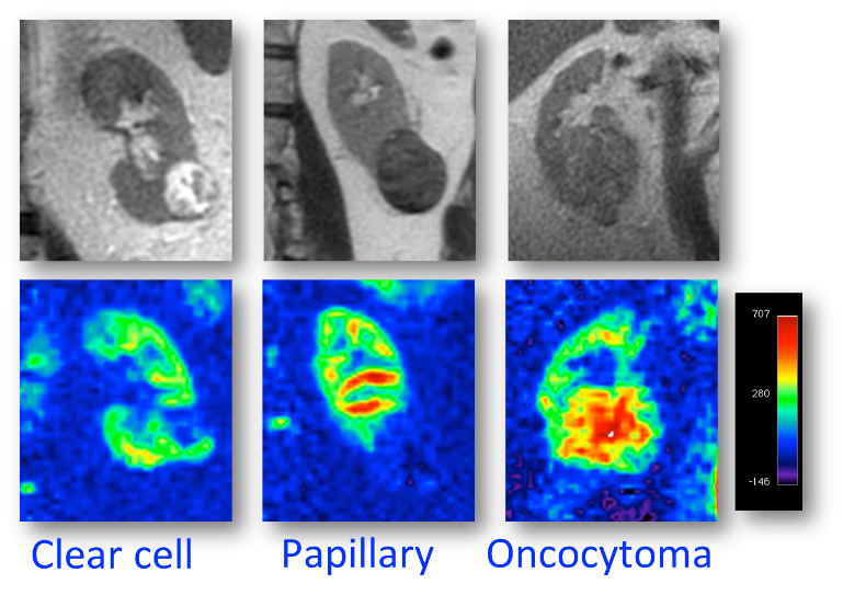
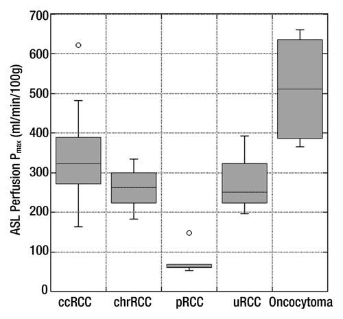
Evaluation of Renal Masses with Arterial Spin Labeled (ASL) MRI: Correlation with Histopathologic Findings
ASL MR imaging enables distinction among different his-topathologic diagnoses in renal masses on the basis of their perfusion level. Oncocytomas demonstrate higher perfusion levels than RCCs, and papillary RCCs exhibit lower perfusion levels than other RCC subtypes.
Lanzman, et al. Radiology 2012