Investigators
- Daniel Costa
- Qing Yuan
- Neil Rofsky
- Ivan Pedrosa
Projects
Diagnostic Performance of Phased-Array only versus endorectal coil multiparametric MRI Protocols for lesion detection
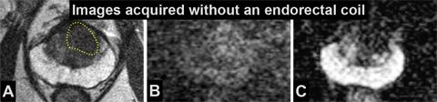
Increased confidence in diagnosis using an endorectal coil (ERC) MR imaging protocol compared to the enhanced protocol. In this 67-year-old man with elevated prostate-specific antigen (9 ng/mL) and one previous negative biopsy, the non-ERC T2-weighted images (A) demonstrated a vague area of homogeneous low signal intensity in the left anterior gland (yellow dotted line) with also subtle increased signal intensity on diffusion-weighted image (B), and low signal intensity on the apparent diffusion coefficient map (C). These findings were prospectively interpreted as equivocal for malignancy (Likert score 3). After insertion of the ERC coil, the T2-weighted images (D) better depict a 23 x 17 mm non-encapsulated mass in the left anterior mid gland with diffusely and homogeneous low-signal intensity (yellow dotted line), obvious increased signal intensity on high b-value diffusion-weighted image (E) and significantly low signal intensity on the ADC map (F) compared to the remainder of the anterior gland; this was interpreted as lesion highly suspicious for malignancy (Likert score 5). A 3- dimensional rendering of the prostate generated with the MR imaging-transrectal ultrasound fusion biopsy (G) reveals positive cores (red) collected from the targeted area (yellow) with Gleason score 3+4 in up to 65% of the core (intermediate risk tumor). The increased signal-to-noise of the ERC protocol allowed for a more confident identification of this clinically significant cancer.

MRI-US Fusion Biopsy for Diagnosis of Prostate Cancer
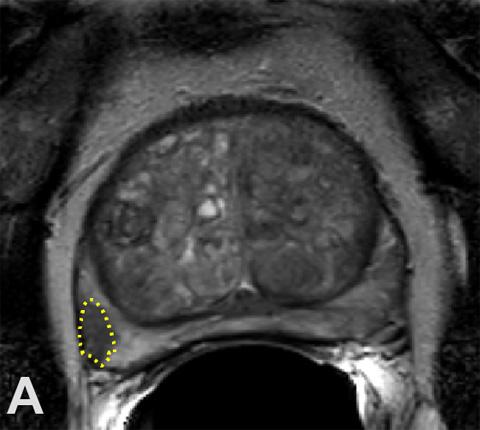
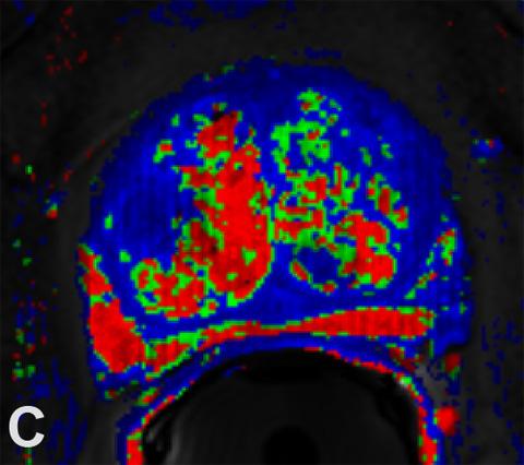
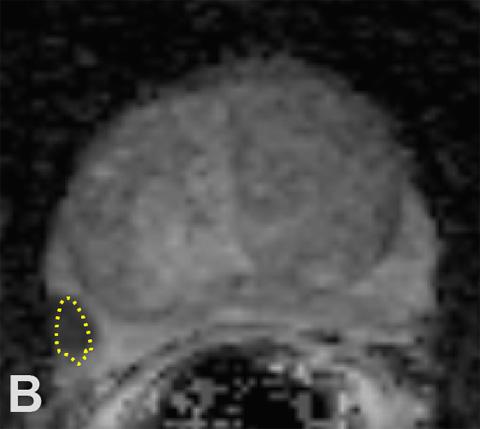
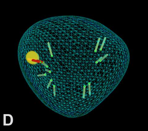
Targeted MRI-TRUS fusion biopsy detecting prostate cancer missed by random biopsies. 67-year-old man with elevated PSA (7 ng/mL) and 2 previous negative biopsies underwent multiparametric MRI for identification of potential targets before repeat biopsy. MRI showed a 1.3 cm nodule in the right lateral mid gland peripheral zone with low signal intensity on axial T2-weighted images (area within dotted line in A), restricted diffusion and low ADC values (B), and abnormal contrast kinetics (red in C) highly suspicious for cancer (Likert scale score of 5). Coronal view of a 3D projection of the prostate generated during the biopsy (D) shows the lesion detected with MRI (yellow), the 2 positive (red) targeted cores revealing Gleason score 3+4 cancer with 50% of maximum core involvement by tumor and the 12 negative (green) systematic cores obtained in the same biopsy session. Radical prostatectomy confirmed a site- and Gleason score-concordant, organ- confined tumor with 1.2 x 0.9 x 0.8 cm.
Performance of Likert Score Scale for Diagnosis of Prostate Cancer
MR-guided HIFU ablation
Collaborators
- Robert Lenkinski
- Rajiv Chopra
- Claus Roehrborn
- Yair Lotan
- Ganesh Raj
- Kevin Courtney