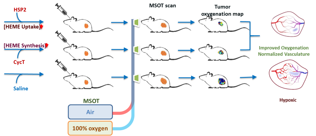
Research

Liu Lab Research

Dr. Liu’s research interests are in development of reporter genes and evaluation different gene reporter applications in pre-clinical cellular and molecular imaging, particularly of small animal models of cancer of breast, prostate, lung and brain, as well as subcutaneous, orthotopic and lung colonization tumors. Using molecular optical imaging techniques, Dr. Liu has conducted evaluations using transfected cell lines and tumor models that investigate the efficacy of several novel technologies such as biodegradable photoluminescent polymers and siRNA delivering functional polymer. Most recently, she explored multispectral optoacoustic tomography (MSOT) to examine the vasculature inside malignant tumors in real time non-invasively, effectively showing that two experimental drugs can normalize aberrant blood vessels, oxygenation, and other aspects of the tumor microenvironment in non-small cell lung cancer (NSCLC), helping to suppress the tumor’s growth and spread.

Dr. Liu’s expertise is in developing and evaluating novel therapeutic drugs utilizing dynamic bioluminescent, fluorescent and MSOT imaging related to vascular disputing agents and heme target drugs.

    Lung Cancer Therapy

    Optimizing Therapeutic Strategies Against Lung Cancer Using Multi-Modality Imaging

    Despite improved early detection and the use of targeted therapies and immunotherapies, the lung cancer 5-year survival rate remains 10-20% in most countries. Tumor hypoxia, i.e., oxygen deprivation, is an independent marker of poor prognosis in many types of human cancer. Substantial tumor hypoxia exists in non-small cell lung cancer (NSCLC), even in early-stage tumors. NSCLC represents 85-90% of all lung cancer cases. Importantly, recent studies in human patients and mice have shown that NSCLC tumors exhibit elevated oxidative metabolism relative to adjacent normal tissues. Our studies also showed that NSCLC cells have intensified oxygen consumption, as well as elevated synthesis and uptake of heme. Heme is a molecule crucial for the functioning of many proteins and enzymes involved in using oxygen to generate cellular energy.

    We have identified two new drugs that inhibit oxidative metabolism and oxygen consumption in NSCLC cells. These two drugs substantially delay lung tumor growth and progression in mouse models of lung cancer. They have a high potential to alleviate tumor hypoxia. Our goal is to illuminate the mechanisms by which the new drugs target oxidative metabolism, alone or in combination with chemotherapy or radiotherapy, influence lung tumor growth, vasculature, and oxygenation. We will characterize the responses of lung tumors in several representative mouse models to the new drugs and combination therapies. We will use advanced imaging techniques, together with histological and immunohistochemistry analyses, to monitor how lung tumors and their microenvironment respond to these drugs. We will determine how pretreatment with these drugs alters the responses of lung tumors and their microenvironment to chemotherapy and radiotherapy. The successful completion of this project should discover novel strategies to alleviate tumor hypoxia and dramatically improve the therapeutic outcome of lung cancer patients.

    Kidney Cancer Therapy MSOT reveals progression of tumor vascular shut-down following administration of the classic VDA CA4P

    Vascular Image-Guided Optimization of Response (VIGOR) to Therapy in Kidney Cancer

    We will develop the application of a promising novel vascular disrupting agent (VDA) in combination with leading therapies to enhance treatment of kidney cancer. Renal cell carcinoma (RCC) is usually characterized by inactivation of the von Hippel Lindau (vHL) tumor suppressor protein, promoting accumulation of Hypoxia Inducible Factor (HIF) and consequent development of extensive vasculature. The endothelium of normal blood vessels is largely quiescent, but the invasive neovasculature of tumors is immature, lacks pericyte support, and exhibits increased permeability providing a selective target for cancer therapy. The therapeutic goal of VDAs is to cause rapid widespread disruption of established tumor vasculature leading to regional ischemia, induction of hypoxia and tumor necrosis.

    We have identified OXi8007 as a new potent, water-soluble VDA prodrug generating protracted vascular disruption, dose dependent tumor growth delay and no apparent systemic toxicity. However, VDA monotherapy generally results in re-growth at the tumor periphery and OXi8007 will likely be most effective in augmenting current lead therapies based on complementary modes of action. We will investigate a small-molecule HIF-2 antagonist, PT2977 developed by Peloton Therapeutics, which represents a new class of chemotherapeutic in early clinical trials. Meanwhile, cabozantinib, the small- molecule kinase inhibitor that targets the c-MET receptor, AXL, and VEGFR-2 was recently approved as a first line treatment option for patients with metastatic RCC. We also recognize the emerging success of immunotherapy and anticipate that OXi8007-induced necrosis will enhance antigen presentation promoting response.

    Our overarching hypothesis is that combining these therapeutic approaches will achieve robust long term control of RCC. Investigations will benefit from the resources of the UT Southwestern Kidney Cancer SPORE, which has developed a number of new patient derived tumor lines exhibiting differential sensitivity to HIF-2 antagonists. Effective therapy combination will likely depend on timing of administration of the respective agents and non- invasive imaging will reveal the spatial and temporal pharmacodynamics of tumor response. Bioluminescence imaging (BLI) will effectively interrogate luciferase-transfected RENCA cells in immunocompetent mice. In addition, recently available multispectral optoacoustic tomography (MSOT) non-invasively reveals vascular extent and regional oxygenation without the need for exogenous reporter molecules or cell transfection. Complementary cell-based studies are designed to further explore OXi8007 mechanism of action. Effectively combining targeted therapies should enhance treatment and ultimately survivorship of kidney cancer patients. The goal of these investigations is to demonstrate effective combination therapy as a foundation for investigations in large animals and translation to the clinic.

    Regulation of breast cancer bone metastasis BCCs induced the BM-intrinsic XBP1s signaling in C57BL/6 mice.

    The interactions between breast cancer cells (BCCs) and bone microenvironment (BM) enhance BCC growth, render them drug resistant, and mediate BCC-induced bone destruction, resulting in increased mortality and compromising life quality of breast cancer patients. Bone marrow stromal cells (BMSCs) are progenitors of skeletal tissue components such as bone, cartilage, the hematopoiesis-supporting stroma, and adipocyte-lineage cells. These cells are considered a key player in the BM support of breast cancer bone metastasis (BCBM). BMSCs secret many chemokines, growth factors, exosomes, and inflammatory cytokines to promote homing, survival and growth of BCCs in BM, induce dormancy and consequent drug resistance of BCCs, and activate osteoclasts, the bone resorbing cells, to induce osteolytic lesions in bone. We reason that BMSCs need to upregulate their capacities of protein folding and trafficking in order to accommodate their increased protein synthesizing and secretion activities.

    The tumor bone microenvironment exhibits heightened inflammation, hypoxia and nutrient-deprivation, all of which are potent inducers to activate ER stress signaling. X-box binding protein spliced (XBP1s) is a basic-region leucine zipper (bzip) transcription factor of the CREB-ATF protein family and a major proximal effector of ER stress signaling. Upon ER stress, Xbp1 mRNA is spliced via an ER-localized endoribonuclease/kinase Inositol Requiring Enzyme-1α (IRE1α) to generate spliced Xbp1 mRNA, termed Xbp1s, from unspliced Xbp1 (Xbp1u). Xbp1s encodes an active transcription factor (XBP1s) that drives gene expression of a wide range of molecules involved in maintenance of intracellular protein homeostasis.

    We discovered that human XBP1s (hXBP1s) is a stromal-intrinsic oncogenic factor that enables BMSC support of multiple myeloma (MM) cell growth and MM-induced bone destruction in multiple myeloma bone disease (MMBD). MM patient BMSCs have a heightened level of protein expression of hXBP1s, compared with normal donor BMSCs. Overexpression of hXBP1s in human BMSCs (hBMSCs), KM101 cells, granted them an array of pathological features of MM BMSCs and thus enhanced stromal support of MM cell growth and osteolysis both in vivo and in vitro. Knocking down hXBP1s in MM BMSCs leads to significant reversal of MM BMSCs’ pathological behaviors. Similar to MMBD, 70% of BCBM are osteolytic. Intriguingly, the BCC-intrinsic XBP1s was recently found to be an important oncogene that renders drug resistance and relapse of triple negative (TN) BCCs. We hypothesize that the stromal IRE1α /XBP1s pathway is also an essential pathogenic factor that potentiates the stromal support of BCC growth and BCC-induced osteolysis, and thus a potential novel therapeutic target for disrupting the stromal support to the progression of BCBM. Upon successful completion, our studies could provide an important rationale for targeting the IRE1α/XBP1s pathway for treating breast cancer. Such a therapeutic strategy could likely achieve an effect of killing two birds with one stone – overcoming drug resistance and relapse of BCCs and disrupting the stromal support to BCBM simultaneously.