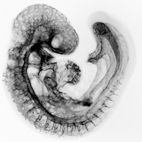
One interesting characteristic of blood vessels that course throughout all tissues and organs is that they are morphologically, transcriptionally, and functionally adapted to their local microenvironment. This heterogeneity can be appreciated by the very nature of the highly hierarchical arrangement of large and small vessels. Arteries and arterioles form closed circulatory loops with veins, via fine networks of capillaries, the smallest blood vessels.
Blood vessels in different tissues must accomplish different functions, and therefore endothelial cells within these tissues take on very different ultrastructural appearances. For instance, organs involved in filtration, such as the pancreas and kidney, possess vessels with pores (fenestrations). Discontinuous capillaries develop much larger pores, or sinusoids, like those in the liver.
These organ-specific endothelial traits are critical for vessels serving organ-specific functions. How and when in the lifespan of an organism do endothelial cells, derived from seemingly similar progenitors, give rise to such heterogeneous populations? Do they emerge de novo distinct from different regions of the mesoderm? Do angioblasts give rise to daughter cells with different fates?
We are carrying out studies that compare different vascular beds across different developmental stages using microarray analysis and RNAseq. The challenge ahead is recognizing those differences in regionally defined vascular beds, understanding how they are molecularly governed and how this could help direct the development of therapeutic applications.
