Primary cilia and compartmentalized signaling
What are primary cilia?
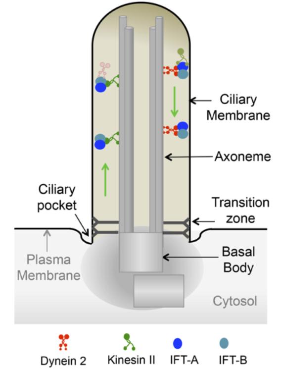
Most cells in our body have an organelle called the primary cilium. The first cellular organelle to be described in biology, the primary cilium was long mistaken as a vestigial appendage. The primary cilia are now considered as vital sensory organelles for detection and transmission of a broad range of chemical and mechanical signals. The non-motile primary cilia are widely present in quiescent cells in our body (except in cells from myeloid and lymphoid lineages) (Rosenbaum and Witman, 2002). Structurally, the primary cilium has a central core of nine microtubule doublets (9+0), which is templated from the mother centriole with associated pericentriolar material (called the basal body) (Figure 2). The assembly and disassembly of the primary cilium is also integrated into the cell cycle, with assembly occurring during the G1/G0 phase, and disassembly before mitosis during the G2 phase (Kobayashi and Dynlacht, 2011). The axoneme is surrounded by the ciliary membrane, which is an extension of the contiguous plasma membrane. A specified region at the base of the cilium (called the transition zone) results in compartmentalization of components in the cilia (Reiter et al., 2012). An adjacent region of the plasma membrane (called the ciliary pocket) is rich in endocytosis components, while primary cilia are typically devoid of endosomes (Rohatgi and Snell, 2010).
Why do the cells require a primary cilium for organizing signaling?
Localization of endogenous proteins in ciliary membrane of vertebrate cells has been mostly determined by immunolabelling techniques. However, it is important to realize that these proteins are not exclusive to cilia; rather, they are enriched in cilia. The ciliary membrane is about 1/1000-1/5000 of the total cellular surface, and the ciliary volume (~0.5 fl) is about ~1/30,000 of the total cellular volume (Delling et al., 2013). The small size of the cilium enables enrichment of proteins with respect to the rest of the plasma membrane, and in establishing an effective signaling compartment by local concentration of second messengers and effectors. However, the absolute amounts of cilia-localized proteins are likely to be minute in comparison to total cellular levels. Thus, to understand the role of compartmentalization in ciliary signaling it is imperative to determine mechanisms underlying ciliary trafficking, and identify functional consequences upon disruption of ciliary localization. Unfortunately, we are currently lacking in our understanding of signaling inside cilia, mostly due to the difficulty of working with such a tiny compartment. We are also extremely limited in availability of tools that allow us to address the role of ciliary compartmentalization while maintaining the architecture of cilia and/or retaining the functionality of the studied proteins (Mukhopadhyay et al., 2017). The main focus on our research is to address the central question of why and how cells utilize these singular organelles as detection and signal-transducing machines.
References
Bhogaraju, S., Engel, B.D., and Lorentzen, E. (2013). Intraflagellar transport complex structure and cargo interactions. Cilia 2, 10.
Delling, M., DeCaen, P.G., Doerner, J.F., Febvay, S., and Clapham, D.E. (2013). Primary cilia are specialized calcium signalling organelles. Nature 504, 311-314.
Hildebrandt, F., Benzing, T., and Katsanis, N. (2011). Ciliopathies. N Engl J Med 364, 1533-1543.
Kobayashi, T., and Dynlacht, B.D. (2011). Regulating the transition from centriole to basal body. J Cell Biol 193, 435-444.
Kozminski, K.G., Johnson, K.A., Forscher, P., and Rosenbaum, J.L. (1993). A motility in the eukaryotic flagellum unrelated to flagellar beating. Proceedings of the National Academy of Sciences of the United States of America 90, 5519-5523.
Mukhopadhyay, S., Badgandi, H.B., Hwang, S.H., Somatilaka, B., Shimada, I.S., and Pal, K. (2017). Trafficking to the primary cilium membrane. Mol Biol Cell 28, 233-239.
Reiter, J.F., Blacque, O.E., and Leroux, M.R. (2012). The base of the cilium: roles for transition fibres and the transition zone in ciliary formation, maintenance and compartmentalization. EMBO reports 13, 608-618.
Rohatgi, R., and Snell, W.J. (2010). The ciliary membrane. Current opinion in cell biology 22, 541-546.
Rosenbaum, J.L., and Witman, G.B. (2002). Intraflagellar transport. Nat Rev Mol Cell Biol 3, 813-825.
Scholey, J.M. (2008). Intraflagellar transport motors in cilia: moving along the cell's antenna. The Journal of cell biology 180, 23-29.